本篇以 Helseth HC, Mansur P, El-Baba M, McLaren JTT, de Alencar JN, Smith SW. Electrocardiographic principles for the diagnosis of occlusion myocardial infarction. Eur Heart J Acute Cardiovasc Care. 2026. DOI: 10.1093/ehjacc/zuag114 為主體。1
這個月讀到一篇文章,我覺得應該讓每個ED man都讀過。
作者群你一看就知道是誰:Stephen Smith、Jesse McLaren、José Nunes de Alencar。OMI這套東西的原班人馬。
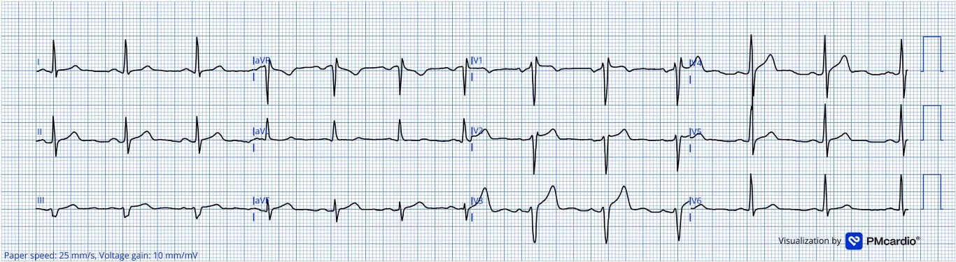
Fig. 1. 貫穿全文的病例,第一張 ECG:40 歲男性,胸痛。下壁與前壁導程的 T 波偏大,但看不出是不是 HATW。原始 ECG 上 V3 的 S 波太深,紙上印不下被切掉了;這張是數位化重繪的版本,所以圖上看不出來。Queen of Hearts 判讀:未偵測到 OMI。圖片出處:Helseth HC, Mansur P, El-Baba M, McLaren JTT, de Alencar JN, Smith SW. Electrocardiographic principles for the diagnosis of occlusion myocardial infarction. Eur Heart J Acute Cardiovasc Care. 2026: Figure 2. DOI: 10.1093/ehjacc/zuag114
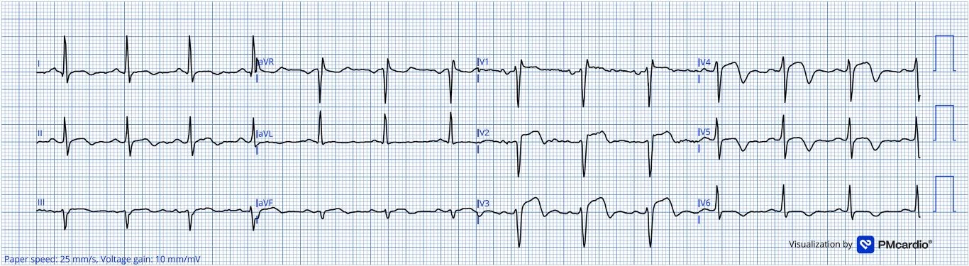
Fig. 2. 同一位病人 16 分鐘後的第二張 ECG。T 波相對於前面的 QRS 明顯不成比例地肥大,合併終末 T 波倒置。作者判讀為前壁與下壁 OMI 合併再灌流,形態介於 active 與 reperfused 之間,病人還痛不痛是判斷的關鍵。Queen of Hearts 判讀:OMI。圖片出處:Helseth HC, Mansur P, El-Baba M, McLaren JTT, de Alencar JN, Smith SW. Electrocardiographic principles for the diagnosis of occlusion myocardial infarction. Eur Heart J Acute Cardiovasc Care. 2026: Figure 3. DOI: 10.1093/ehjacc/zuag114
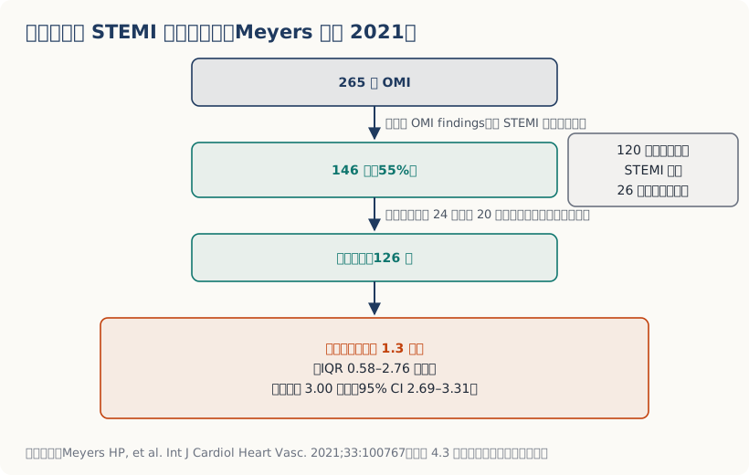
① 有一群人,你等再久它都不會達到門檻。 Meyers等人2025年把53例LAD完全阻塞(TIMI-0,完全沒有血流)的病人,血管攝影前的每一張ECG都拿來量STEMI準則,結果有20例(38%)「任何一張」都沒有達到。這20例裡有16例做了兩張以上,第一張到最後一張中位數隔了44分鐘(最久隔了44小時),還是沒有一張達到。7
③ 通了也可能再塞回去。 Lemkes等人2019年那個transient STEMI的RCT,延遲介入組有5.6%(4例)因為再梗塞的症狀與徵象而需要緊急介入。9
症狀好轉、ECG變漂亮,不等於問題解決了。那可能只是血栓暫時鬆開。
所以在急診你要怎麼做?
症狀有變化 ➜ 重做ECG。 不是等下一個班,是現在。
第一張non-diagnostic但你心裡毛毛的 ➜ 設定時間點重做。 那篇文章的貫穿病例是16分鐘。 那到底該多久做一張?這個數字是有出處的,而且它有一段演變。 2014年的AHA/ACC NSTE-ACS指引寫得很白:10「The ECG can be relatively normal or initially nondiagnostic; if this is the case, the ECG should be repeated (e.g., at 15- to 30-minute intervals during the first hour), especially if symptoms recur.」 講成中文就是:最初一小時內,每15到30分鐘一張。
但2025年的新版把這個固定間隔拿掉了。 這份新版是2025 ACC/AHA/ACEP/NAEMSP/SCAI ACS指引,把STEMI和NSTE-ACS合在一起寫,改成「依症狀與臨床狀態的變化重做」(原文:「timing of repeat ECGs should be guided by the patient’s symptoms, especially recurrent chest pain, and any change in clinical condition」)。11
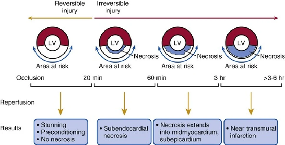
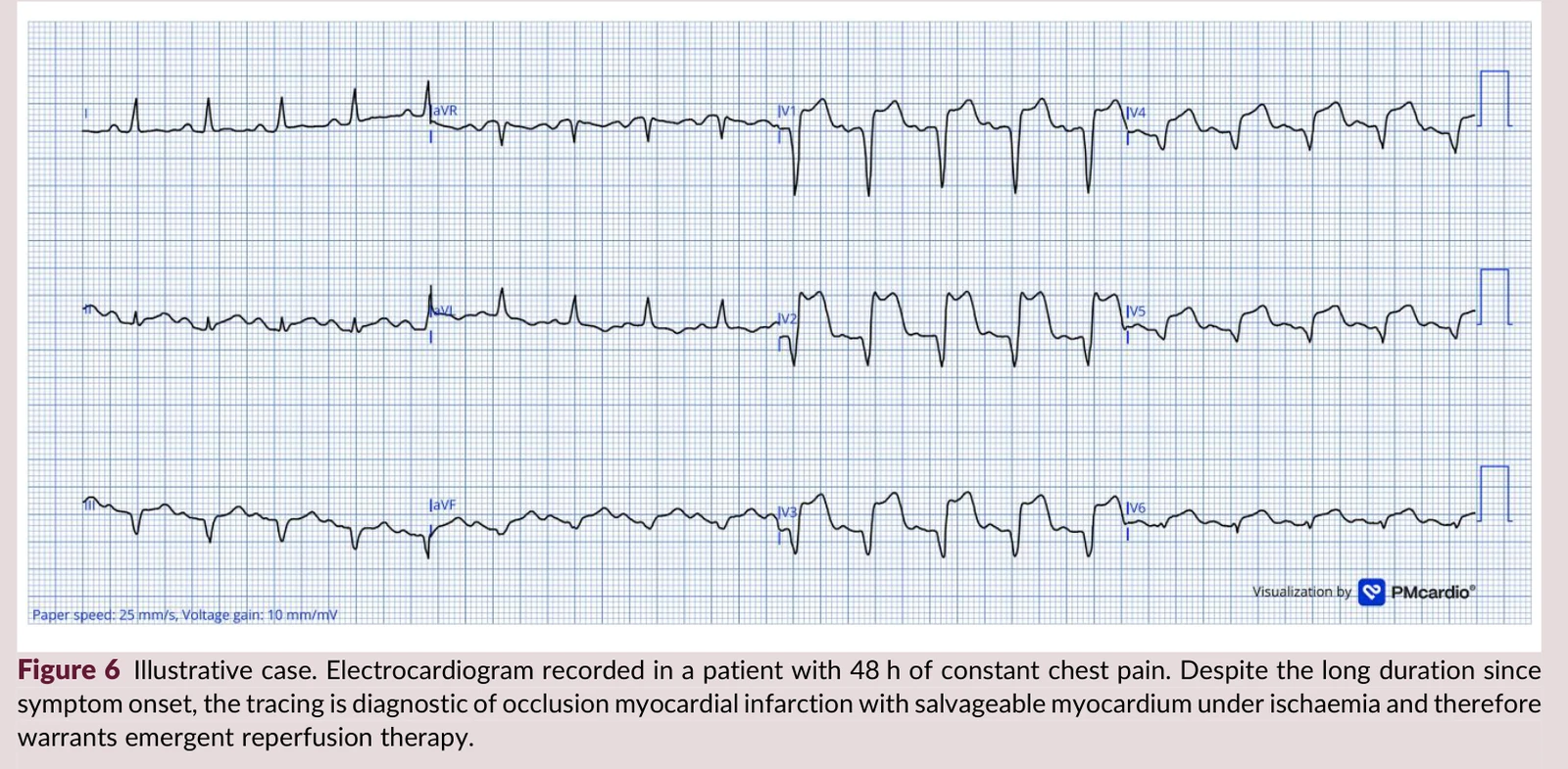
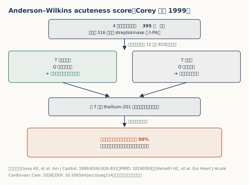
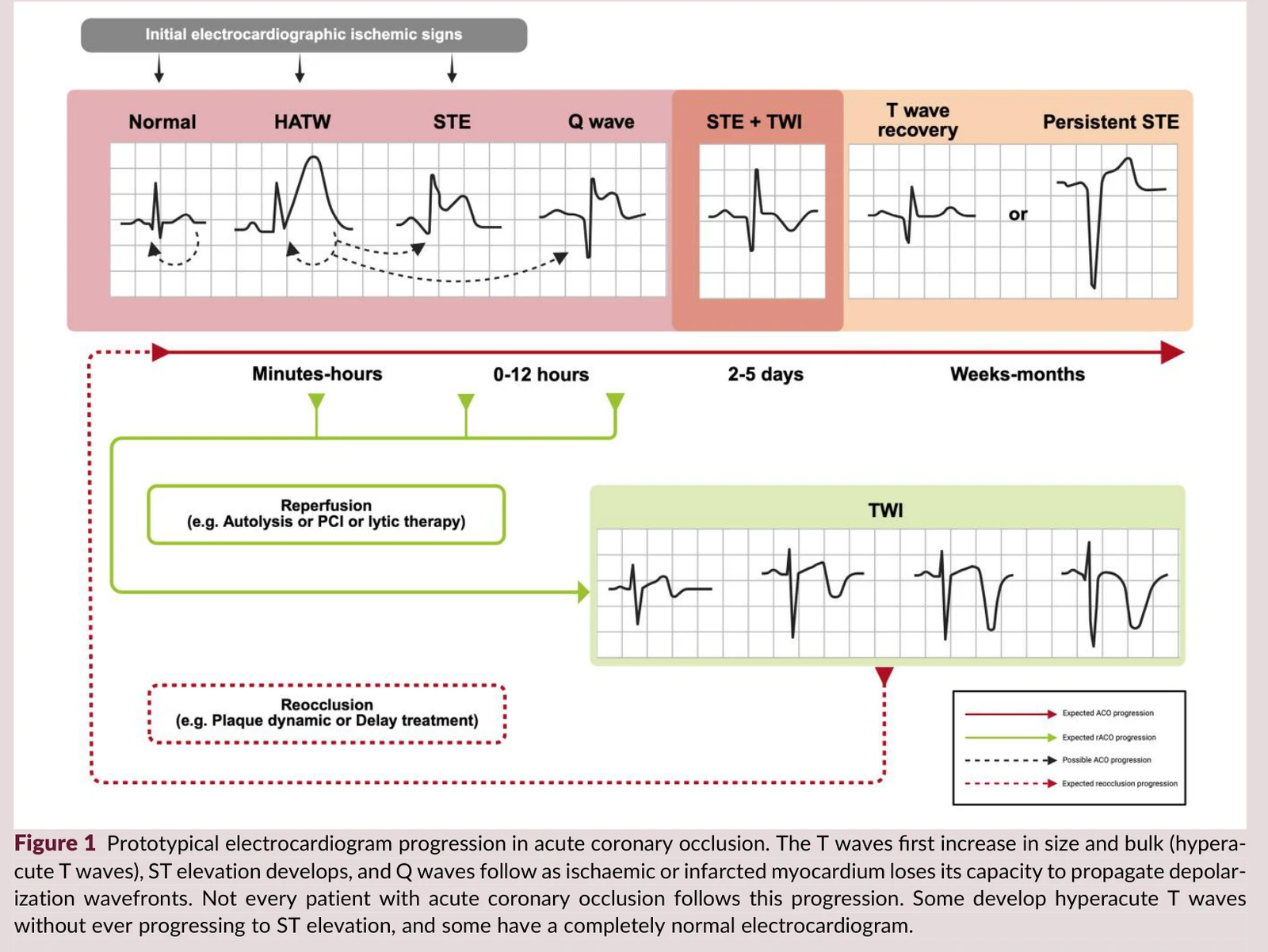
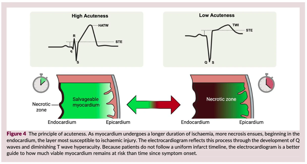
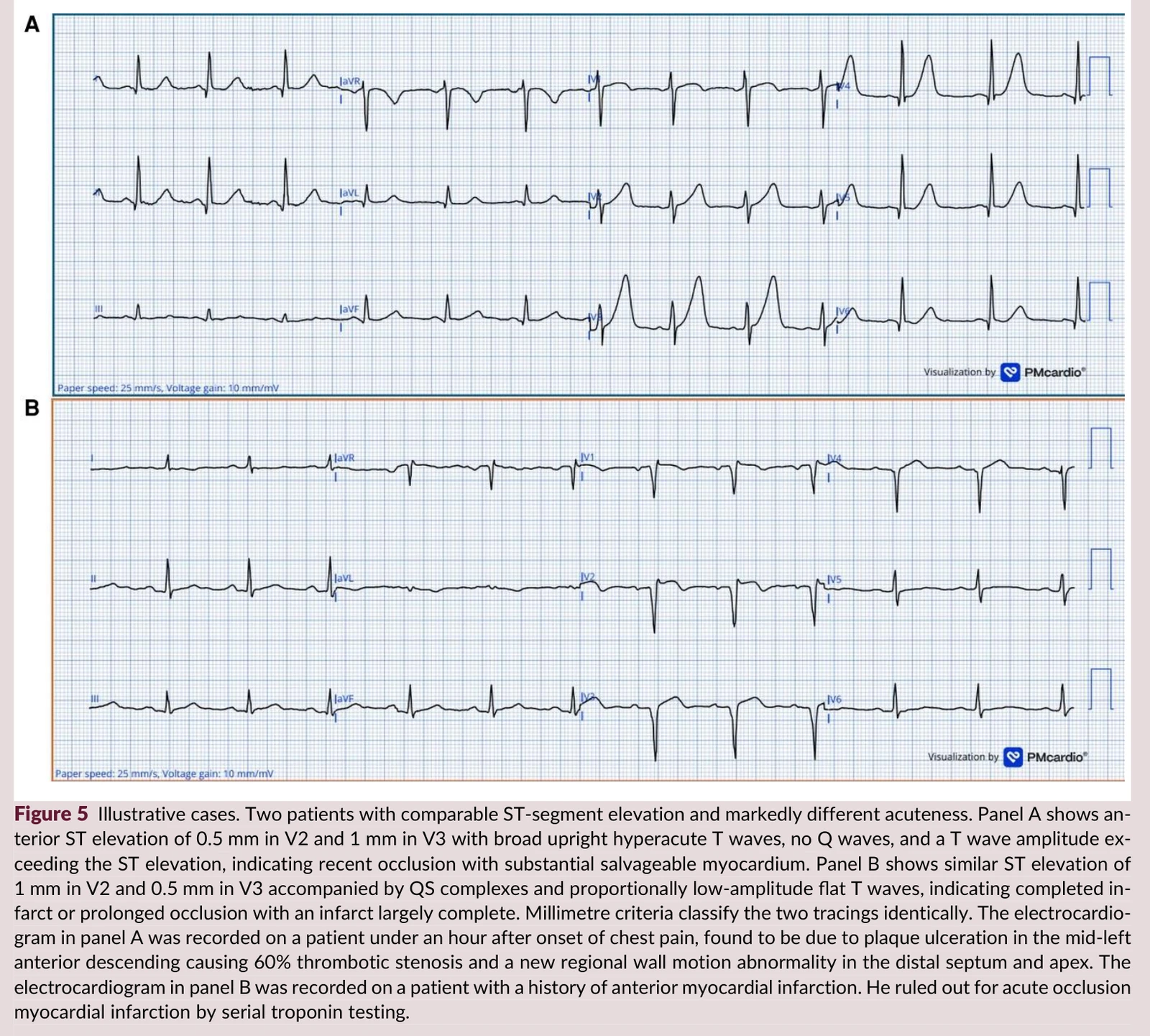
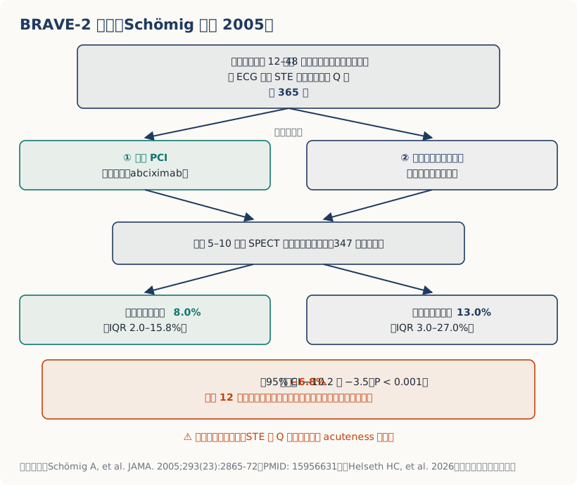
原文在acuteness這一節寫了一句:超過傳統的時間窗,只要acuteness高,緊急PCI還是有好處(原文:「Beyond traditional time windows, emergent PCI remains beneficial when acuteness is high」),然後拿BRAVE-2當這句話的證據。
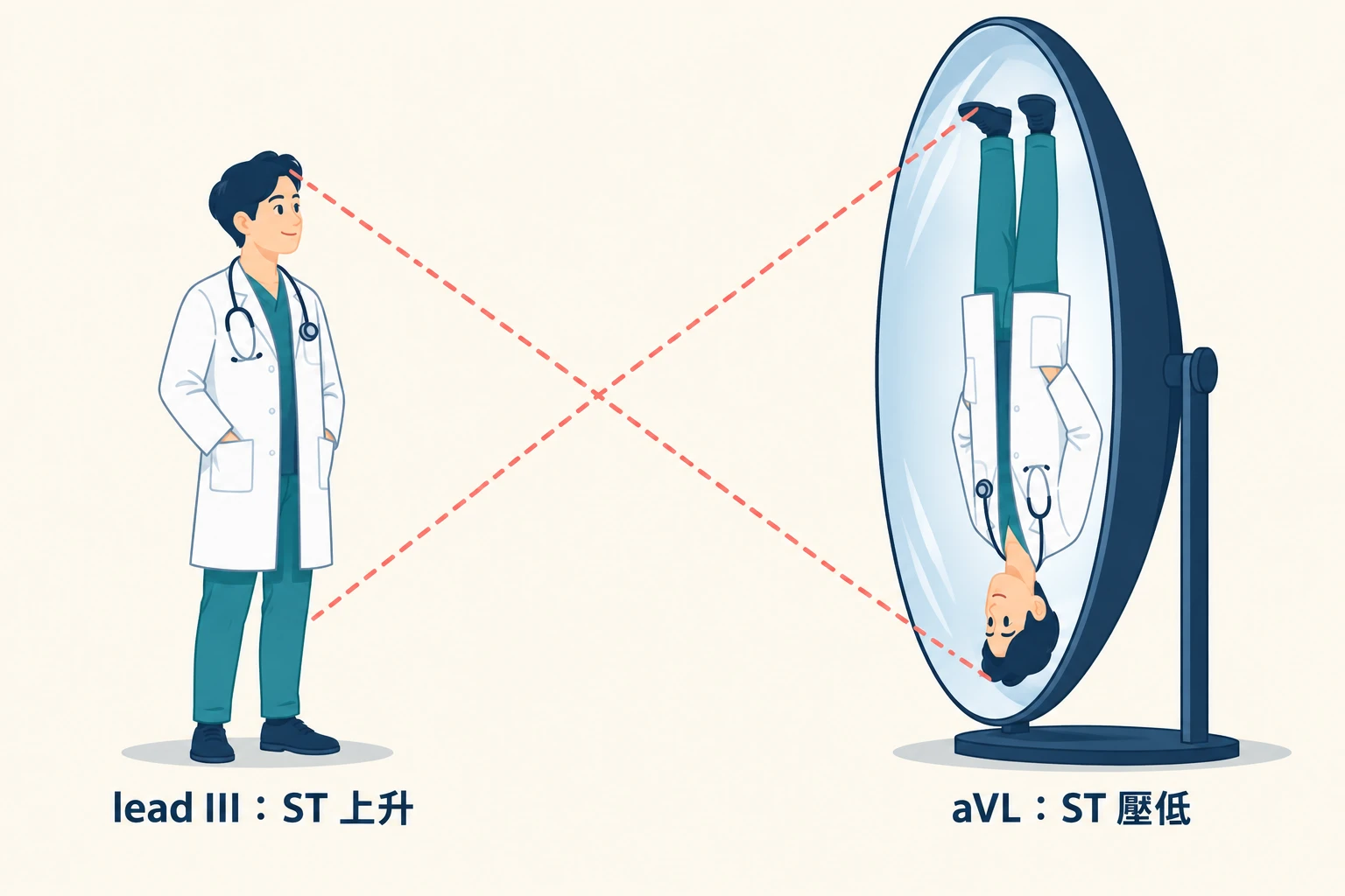
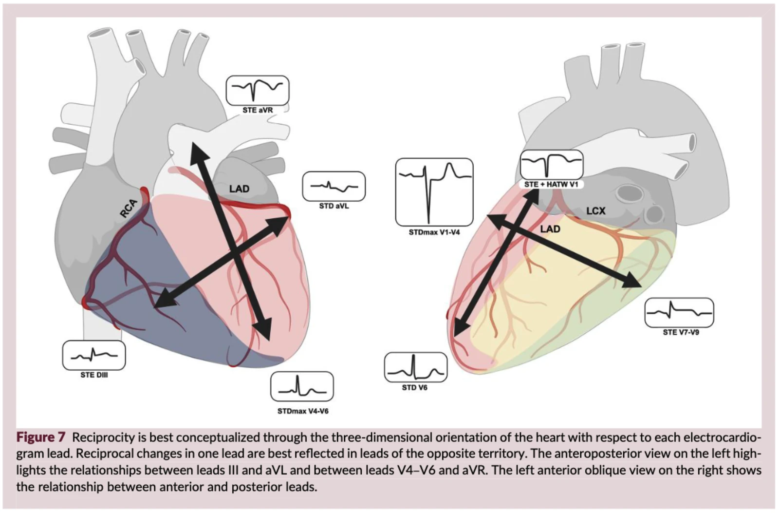
講到這裡,一定要提 Dr. Jerry Jones 對 reciprocal change 的兩個提醒。他的原話是: "Reciprocal changes to an acute occlusion of one of the coronary arteries may appear before any ST elevation. And even if the ST elevation is present, the reciprocal changes may continue to be much much more impressive. Don't be fooled." 翻成白話文就是兩件事:
① reciprocal change(STD)可能比STE更早出現。 你還在等STE的時候,鏡子那一邊可能已經先講話了。
② 就算STE已經出現,STD還是可能比STE更明顯。 所以不要因為「STE看起來不怎麼樣」就放過它。
還有一個更麻煩的:Geffin等人2024年指出,left dominant合併近端迴旋支阻塞時,高側壁與下壁的向量可能大致互相抵銷。這就是為什麼隱性(silent)OMI最常發生在迴旋支區域。原文逐字是:「In left dominant circulation with proximal circumflex occlusion, high lateral and inferior vectors may roughly oppose each other, which is why silent OMI most often involves the circumflex territory.」同一段還補了一句:left main阻塞同時造成前壁與後壁OMI的時候,抵銷得更不均勻。
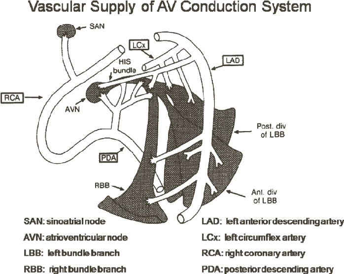
① 節律與傳導:血管會告訴你它塞在哪Fig. 16. 房室傳導系統的血液供應:RCA 供應 AV node 與 His 束近端;LAD 的中膈穿通支供應 His 束遠端、右束支與左前分支。圖片出處:Hsing JM, Hsia HH. Cardiac Arrhythmias. In: Criner GJ, Barnette RE, D’Alonzo GE, eds. Critical Care Study Guide: Text and Review. 2nd ed. Springer; 2010: Figure 19-3(modified from DeGuzman)
講到這裡,順便自己推銷一下。
2024年我寫過一篇 How to detect OMI in 10 Steps?,把「怎麼在一張ECG上一步一步找OMI」拆成十個步驟,從排除artifact開始,一路走到各種STE/STD的情境。
那篇是操作手冊,這篇是心法。
六條原則講的是「為什麼要這樣看」,十個步驟講的是「實際上手怎麼看」。兩篇搭著讀,效果會比較好。
學習重點:
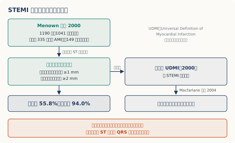
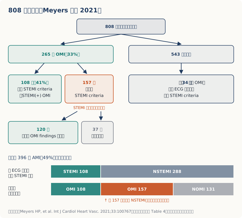
STEMI毫米門檻不是用血管攝影當Gold Standard推導出來的,也沒把比例性納入。 所以它對ACO的敏感度只有43.6%(95% CI 34.7–52.9%)。沒塞住的人它幾乎不會誤判,真的塞住的人它有一半以上抓不到。
Surrogacy:ECG是證人,不是法官。 NSTEMI病人中25.5%罪犯血管是塞的,死亡RR 1.67(95% CI 1.31–2.13);而疑似STEMI的人裡,16%在血管攝影時已經自己通了。ECG是用來修正機率的,不是用來定義阻塞的。AI也一樣:演算法跟你一樣,只看得到心電圖,看不到冠狀動脈。
Helseth HC, Mansur P, El-Baba M, McLaren JTT, de Alencar JN, Smith SW. Electrocardiographic principles for the diagnosis of occlusion myocardial infarction. Eur Heart J Acute Cardiovasc Care. 2026. DOI: 10.1093/ehjacc/zuag114 ↩︎
Menown IB, Mackenzie G, Adgey AA. Optimizing the initial 12-lead electrocardiographic diagnosis of acute myocardial infarction. Eur Heart J. 2000;21(4):275-283. PMID: 10653675. DOI: 10.1053/euhj.1999.1748;The Joint European Society of Cardiology/American College of Cardiology Committee. Myocardial infarction redefined—a consensus document of the Joint European Society of Cardiology/American College of Cardiology Committee for the Redefinition of Myocardial Infarction. Eur Heart J. 2000;21(18):1502-1513. PMID: 10973764/J Am Coll Cardiol. 2000;36(3):959-969. PMID: 10987628 ↩︎
Macfarlane PW, Browne D, Devine B, et al. Modification of ACC/ESC criteria for acute myocardial infarction. J Electrocardiol. 2004;37 Suppl:98-103. PMID: 15534817. DOI: 10.1016/j.jelectrocard.2004.08.032 ↩︎
Thygesen K, Alpert JS, White HD. Universal definition of myocardial infarction. J Am Coll Cardiol. 2007;50(22):2173-2195. PMID: 18036459(Table 3);Wagner GS, Macfarlane P, Wellens H, et al. AHA/ACCF/HRS recommendations for the standardization and interpretation of the electrocardiogram: part VI: acute ischemia/infarction. J Am Coll Cardiol. 2009;53(11):1003-1011. PMID: 19281933;Thygesen K, Alpert JS, Jaffe AS, et al. Third universal definition of myocardial infarction. J Am Coll Cardiol. 2012;60(16):1581-1598. PMID: 22958960(Table 3);Thygesen K, Alpert JS, Jaffe AS, et al. Fourth Universal Definition of Myocardial Infarction (2018). Circulation. 2018;138(20):e618-e651. PMID: 30571511(Table 2);Mills NL, Newby LK, Zaman S, et al. Fifth Universal Definition of Myocardial Infarction (2026). Circulation. 2026. DOI: 10.1161/CIR.0000000000001477(§13、Table 5) ↩︎
de Alencar Neto JN, et al. Systematic review and meta-analysis of diagnostic test accuracy of ST-segment elevation for acute coronary occlusion. Int J Cardiol. 2024;402:131889. PMID: 38382857. DOI: 10.1016/j.ijcard.2024.131889 ↩︎
Strauss DG, Schocken DD. Marriott's Practical Electrocardiography. 13th ed. Wolters Kluwer; 2021: Chapter 6(Introduction to Myocardial Ischemia and Infarction);Goldberger AL, Goldberger ZD, Shvilkin A. Goldberger's Clinical Electrocardiography: A Simplified Approach. 9th ed. Elsevier; 2018: Chapter 10 ↩︎
Meyers HP, Sharkey SW, Herman R, de Alencar JN, Shroff GR, Frick WH, Smith SW. Failure of standard contemporary ST-elevation myocardial infarction electrocardiogram criteria to reliably identify acute occlusion of the left anterior descending coronary artery. Eur Heart J Acute Cardiovasc Care. 2025;14(7):403-411. PMID: 40717627. DOI: 10.1093/ehjacc/zuaf037 ↩︎
Meyers HP, Bracey A, Lee D, Lichtenheld A, Li WJ, Singer DD, et al. Accuracy of OMI ECG findings versus STEMI criteria for diagnosis of acute coronary occlusion myocardial infarction. Int J Cardiol Heart Vasc. 2021;33:100767. PMID: 33912650. PMCID: PMC8065286. DOI: 10.1016/j.ijcha.2021.100767 ↩︎↩︎↩︎↩︎
Lemkes JS, Janssens GN, van der Hoeven NW, et al. Timing of revascularization in patients with transient ST-segment elevation myocardial infarction: a randomized clinical trial. Eur Heart J. 2019;40(3):283-291. PMID: 30371767. DOI: 10.1093/eurheartj/ehy651 ↩︎
Amsterdam EA, Wenger NK, Brindis RG, et al. 2014 AHA/ACC Guideline for the Management of Patients with Non-ST-Elevation Acute Coronary Syndromes. J Am Coll Cardiol. 2014;64(24):e139-e228. PMID: 25260718. DOI: 10.1016/j.jacc.2014.09.017 ↩︎
Rao SV, O’Donoghue ML, Ruel M, et al. 2025 ACC/AHA/ACEP/NAEMSP/SCAI Guideline for the Management of Patients With Acute Coronary Syndromes. J Am Coll Cardiol. 2025;85(22):2135-2237. PMID: 40013746. DOI: 10.1016/j.jacc.2024.11.009 ↩︎
Riley RF, Newby LK, Don CW, et al. Diagnostic time course, treatment, and in-hospital outcomes for patients with ST-segment elevation myocardial infarction presenting with nondiagnostic initial electrocardiogram: a report from the American Heart Association Mission: Lifeline program. Am Heart J. 2013;165(1):50-56. PMID: 23237133. PMCID: PMC3523309. DOI: 10.1016/j.ahj.2012.10.027 ↩︎
Raitt MH, Maynard C, Wagner GS, Cerqueira MD, Selvester RH, Weaver WD. Appearance of abnormal Q waves early in the course of acute myocardial infarction: implications for efficacy of thrombolytic therapy. J Am Coll Cardiol. 1995;25(5):1084-8. PMID: 7897120 ↩︎
Smith SW, Zvosec DL, Sharkey SW, Henry TD, eds. The ECG in Acute MI: An Evidence-Based Manual of Reperfusion Therapy. Philadelphia: Lippincott Williams & Wilkins; 2002: Chapter 11(Q Waves), p. 96-97 ↩︎
Corey KE, Maynard C, Pahlm O, et al. Combined historical and electrocardiographic timing of acute anterior and inferior myocardial infarcts for prediction of reperfusion achievable size limitation. Am J Cardiol. 1999;83(6):826-831. PMID: 10190393 ↩︎
Schömig A, Mehilli J, Antoniucci D, et al. Mechanical reperfusion in patients with acute myocardial infarction presenting more than 12 hours from symptom onset: a randomized controlled trial (BRAVE-2). JAMA. 2005;293(23):2865-72. PMID: 15956631 ↩︎
Wereski R, Kimenai DM, Taggart C, et al. Cardiac Troponin Thresholds and Kinetics to Differentiate Myocardial Injury and Myocardial Infarction. JAMA Cardiol. 2020;5(11):1302-1304. PMCID: PMC7675101. DOI: 10.1001/jamacardio.2020.2867 ↩︎
Bischof JE, Worrall C, Thompson P, Marti D, Smith SW. ST depression in lead aVL differentiates inferior ST-elevation myocardial infarction from pericarditis. Am J Emerg Med. 2016;34(2):149-154. PMID: 26542793. DOI: 10.1016/j.ajem.2015.09.035 ↩︎
Meyers HP, Bracey A, Lee D, et al. Ischemic ST-Segment Depression Maximal in V1-V4 (Versus V5-V6) of Any Amplitude Is Specific for Occlusion Myocardial Infarction. J Am Heart Assoc. 2021;10(23):e022866. PMID: 34775811. PMCID: PMC9075358. DOI: 10.1161/JAHA.121.022866 ↩︎
Aslanger E, Yıldırımtürk Ö, Şimşek B, et al. DIagnostic accuracy oF electrocardiogram for acute coronary OCClUsion resuLTing in myocardial infarction (DIFOCCULT Study). Int J Cardiol Heart Vasc. 2020;30:100603. ↩︎
Smith SW, Dodd KW, Henry TD, Dvorak DM, Pearce LA. Diagnosis of ST-elevation myocardial infarction in the presence of left bundle branch block with the ST-elevation to S-wave ratio in a modified Sgarbossa rule. Ann Emerg Med. 2012;60(6):766-76. PMID: 22939607. DOI: 10.1016/j.annemergmed.2012.07.119 ↩︎
Klein LR, Shroff GR, Beeman W, Smith SW. Electrocardiographic criteria to differentiate acute anterior ST-elevation myocardial infarction from left ventricular aneurysm. Am J Emerg Med. 2015;33(6):786-90. PMID: 25862248. DOI: 10.1016/j.ajem.2015.03.044 ↩︎
Meyers HP, Simančík F, Herman R, et al. Hyperacute T Waves Are Specific for Occlusion Myocardial Infarction, Even Without Diagnostic ST-Segment Elevation. JACC Adv. 2025;4(10 Pt 2):102120. PMID: 40892623. PMCID: PMC12791876. DOI: 10.1016/j.jacadv.2025.102120 ↩︎
Moon JCC, De Arenaza DP, Elkington AG, et al. The pathologic basis of Q-wave and non-Q-wave myocardial infarction: a cardiovascular magnetic resonance study. J Am Coll Cardiol. 2004;44(3):554-560. PMID: 15358019 ↩︎
Smith SW. Upwardly concave ST segment morphology is common in acute left anterior descending coronary occlusion. J Emerg Med. 2006;31(1):69-77. PMID: 16798159. DOI: 10.1016/j.jemermed.2005.09.008 ↩︎
Stone GW, Cox D, Garcia E, et al. Normal flow (TIMI-3) before mechanical reperfusion therapy is an independent determinant of survival in acute myocardial infarction: analysis from the primary angioplasty in myocardial infarction trials. Circulation. 2001;104(6):636-41. PMID: 11489767 ↩︎
Koyama Y, Hansen PS, Hanratty CG, Nelson GI, Rasmussen HH. Prevalence of coronary occlusion and outcome of an immediate invasive strategy in suspected acute myocardial infarction with and without ST-segment elevation. Am J Cardiol. 2002;90(6):579-84. PMID: 12231080 ↩︎
Khan AR, Golwala H, Tripathi A, et al. Impact of total occlusion of culprit artery in acute non-ST elevation myocardial infarction: a systematic review and meta-analysis. Eur Heart J. 2017;38(41):3082-3089. PMID: 29020244. DOI: 10.1093/eurheartj/ehx418 ↩︎
Herman R, Meyers HP, Smith SW, et al. International evaluation of an artificial intelligence-powered ECG model detecting acute coronary occlusion myocardial infarction. Eur Heart J Digit Health. 2024;5(2):123-133. PMID: 38505483. PMCID: PMC10944682. DOI: 10.1093/ehjdh/ztad074 ↩︎
Herman R, Mumma BE, Hoyne JD, Cooper BL, Johnson NP, Kisova T, et al. AI-Enabled ECG Analysis Improves Diagnostic Accuracy and Reduces False STEMI Activations: A Multicenter U.S. Registry. JACC Cardiovasc Interv. 2026;19(2):145-156. PMID: 41158088. DOI: 10.1016/j.jcin.2025.10.018 ↩︎



看完有什麼想法?在下面留言、按讚或給個表情,一起討論。
comments powered by Disqus