Translated from the original Traditional Chinese post. Read the Chinese original →
Which lead is the most worrying?

A 58-year-old man had just been released from prison and sent home on the day he got sick. That afternoon, his wife found him collapsed in the bathroom, unconscious.
He gradually came around, with crushing chest pain.
Plots like this are strange. Sometimes joy turns to sorrow; sometimes tragedy piles on tragedy.
One I remember from my residency: a senior attending from one of the hospital's departments had retired the day before, and the very next day, while withdrawing money at the bank inside the hospital, collapsed....... a Zhudong glass-seller (Aortic dissection; our ED slang that sounds like the Chinese term).
I've also had a case where we were resuscitating an MI patient in the resus room, and when a family member arrived and heard their relative was having an MI, they collapsed too, with left-sided hemiplegia. The resus room instantly gained one more within-three-hours stroke patient. (lll  ̄v ̄)
Anyway, nobody can predict what will happen in the next moment of life. Don't build a procrastination habit: I'll do it tomorrow, I'll take a look next week...... can you imagine these emergencies filling up your appointment slots before your procrastination even kicks in? OK, that was me talking to myself; this month's post is written.
Back to this case. This was a colleague's case, five minutes before the end of my shift. After the ECG, my colleague decided to call the CV man. Before the nurse practitioner picked up the phone to call tonight's unlucky on-call, they put the ECG in front of me and let me take a look too.
What problems did I see?
RAD
Sinus tachycardia
PRWP(QS wave in V1-V4)
STE in V2-V6, I/aVL with HATW
STD in III/aVF
Let's ask ourselves a few questions first
- How common are RAD and sinus tachycardia in AMI patients?
- With deep QS waves already present in V1-V4, what other conditions need to be considered as well?
- What proportion of ant. wall OMIs show reciprocal STD?
How common are RAD and sinus tachycardia in AMI patients?
I remember once asking medical students: if a patient says they have chest pain, and there are two ECG rates, 80 bpm and 120 bpm, which patient do you think has the higher probability of AMI?
Usually, when an AMI patient is tachycardic, it means they may already be in cardiogenic shock. The heart starts compensating by beating faster. That's why tachycardia appears.
RAD is also uncommon in AMI patients. But if there's a lateral wall MI, you do need to consider it (see Fig.2).
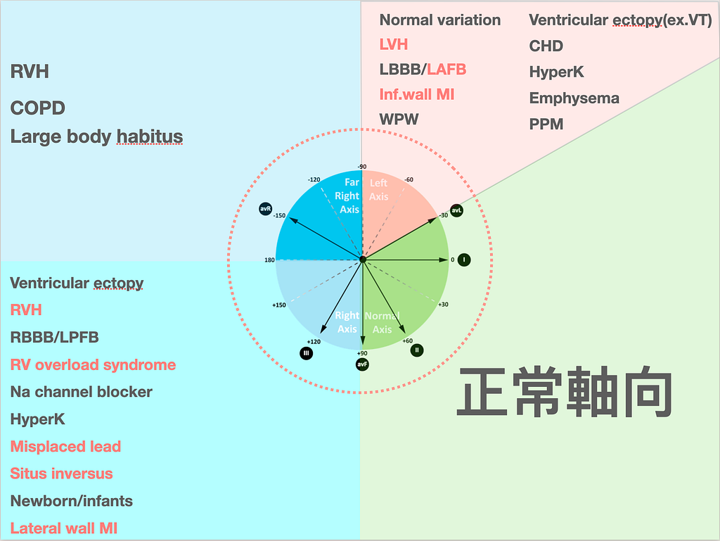
In the DDx list for RAD (lower left corner of Fig.2), besides watching for the things we in the ED care more about, like RV overload syndrome (meaning possible pulmonary embolism), Na channel blocker toxicity, and whether the leads were misplaced, there's also possible lateral wall MI. Also, in kids, because the RV starts out thicker during heart development and only later does the LV become thicker, you may see RAD on ECGs in little kids too~~
My own way of remembering it: the lateral wall sits more toward the outside, so after an AMI, when that myocardium dies, the axis swings to the right (the R wave in lead I gets smaller → somewhat like the concept of PRWP in the precordial leads from AMI)
The DDx list for LAD has one too, and that's inf. wall MI. My way of remembering it: the inf. wall sits even more toward the right side of the heart than the lateral wall, so with an inf. wall MI the axis swings left, producing LAD.
Amal Mattu, in his ECG Weekly teaching, has mentioned many times that STEMI patients rarely have RAD; if you're interested, see this teaching session1.
With deep QS waves already present in V1-V4, what other conditions need to be considered as well?
When there are QS waves in V1-V4, you have to consider whether there was a previous AMI that produced an LV aneurysm. And the ECG pattern of an LV aneurysm already shows STE to begin with.
So how do you tell LV aneurysm STE from the STE of a real AMI?
If you're interested, there are two articles to read.
Derivation Study: published in 20052
Validation Study: published in 20153
Bottom line first: if any one lead in V1-V4 has a T/QRS ratio >0.36, acute STEMI is the more likely diagnosis
Before we get into how to apply the conclusion of these two articles, we need to understand the following questions first; only then can we apply it.
- What conditions does this rule require?
- Which LV aneurysm ECG findings should be put in the DDx?
- How is the rule applied?
Let me answer these one by one~~~
The precondition for this rule is of course that there's STE, and it's used to distinguish acute STEMI from old MI with persistent STE. Also, this rule can't be used for early repolarization vs. STEMI (for that you should use the Smith 4 variable formula)
— — — — — — — — — — Intermission divider — — — — — — — — — —
Here's a quick rundown of the exclusion criteria for the Smith 4 variable formula (Don't know what this thing is? Uh~~~~ when I get a case, I'll write a few more posts to explain it)
- >5 mm STE → exclude
- Non-concave STE → if straight or convex upward STE → exclude
- Inferior reciprocal changes → if there's reciprocal change → exclude
- Anterior ST depression → means there may be reciprocal change → exclude
- Terminal QRS distortion in V2 or V3 → if you see TQRSD, exclude
- Q waves in any of V2 to V4 (means there may already have been an AMI) → exclude
- Any T wave inversion from V2 to V6 (means a possible AMI that then reperfused) → exclude
- BBB present
- One thing you absolutely must watch for → this formula (meaning the Smith 4 variable formula) can't be used in patients with LVH or LV aneurysm; it may give a false-positive
— — — — — — — — — — Intermission divider — — — — — — — — — —
Back to the second question. Which LV aneurysm ECG findings can this rule be applied to?
When there's a well-formed Q wave (at least one lead in V1-V4 has a QS wave)
- QS wave → means no R wave at all, or an r wave of less than 1 mm
The third question is how to apply the rule. As the conclusion above already said: if at least one lead in V1-V4 has a QS wave, then if any one lead (V1-V4) has a T/QRS ratio >0.36, that favors STEMI.
This rule has been checked in a validation study, and this cutoff point has decent sensitivity and specificity
- Derivation study2: showed the accuracy of this formula was 93.2%
- Validation study3: showed sensitivity: 91%, specificity: 81%
Watch out: if chest pain has lasted more than 6 hours, the patient may have a subacute STEMI, so when you apply T/QRS, because so much time has passed the T wave isn't as tall and wide, and it gets classified as LV aneurysm, causing a false-negative.
OK, next, a quick word on the third question from the start
What proportion of ant. wall OMIs show reciprocal STD?
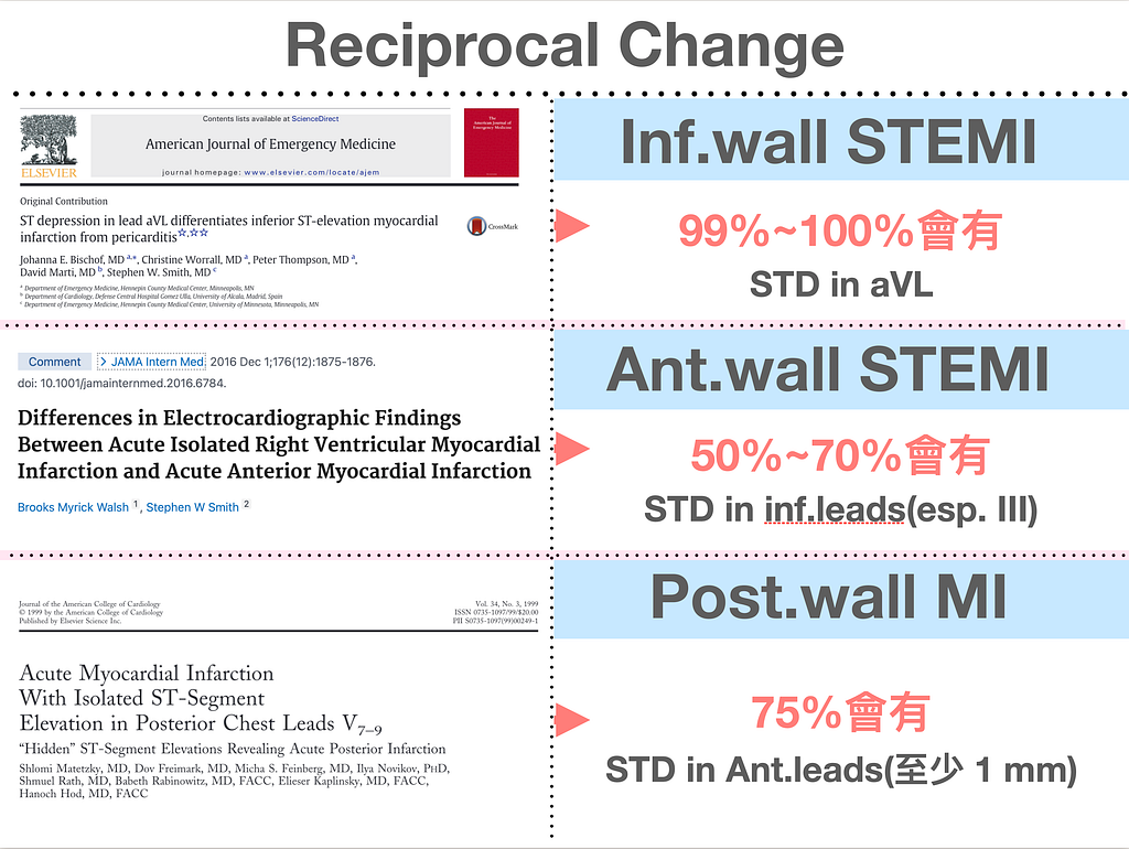
Let's look at how often the opposite leads show reciprocal STD change when there's STE (Fig.3)
- In inf. wall STEMI, 99%~100% have STD in aVL
- In ant. wall STEMI, 50%~70% show STD in the inf.leads (especially III) → usually reciprocal STD change in the inf.leads only shows up when I/aVL are involved (as I said in an earlier post, reciprocal STD change only appears within the same plane)
- In post. wall MI, 75% have STD in the ant.leads (at least 1 mm)
There's one thing I have to stress here: in ant. wall STEMI, the absence of reciprocal STD change in the inf.leads does not rule out ant. wall STEMI; after all, 30%~50% won't show it.
Back to this case~~~
When I saw this ECG in the moment, the leads that struck me at first glance as most worrying weren't V2–V3. Although V2-V3 have very high STE, the QS waves are very deep, so they clearly don't reach a T/QRS ratio greater than 0.36. Instead, V4's T/QRS ratio, eyeballed, should be way over 0.36. (see Fig.4)
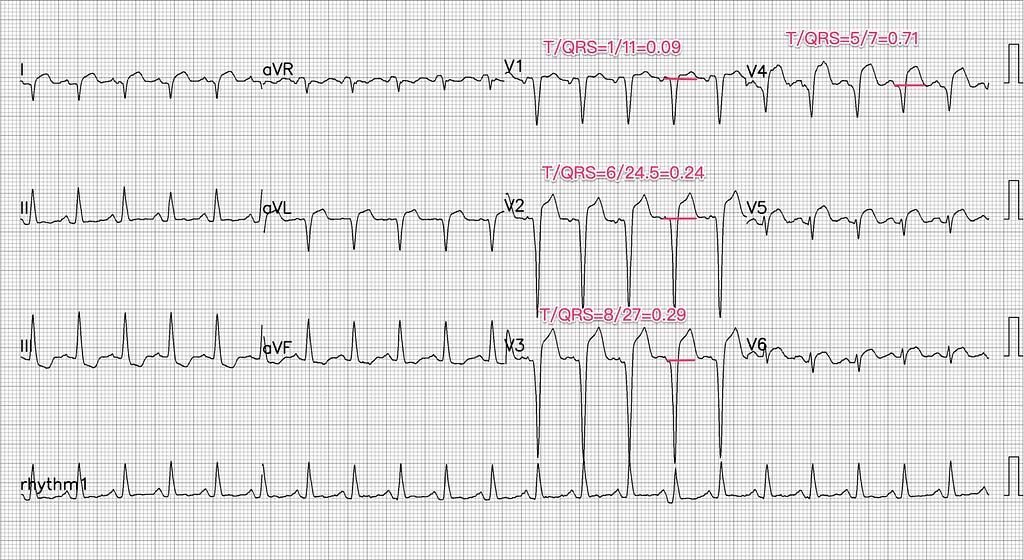
On top of that, V5-V6 also clearly have STE and HATW, with a convex ST segment. I/aVL also have STE, with a straight ST segment shape. These are all obvious ischemic patterns.
III/aVF also show reciprocal STD change.
Later the patient was wheeled into the resus room, and I went to take a look.
No exaggeration: every drop of cold sweat on the patient's forehead was huge. Crushing chest pain. And he kept writhing around, wanting to move his bowels.
O~~~~~M~~~~~G~~~~
My ED brothers and sisters, when you have a critically ill patient on the gurney who keeps wanting to move their bowels, I think the shadow over your heart has got to be enormous~~~
What I often see is that before long, the patient starts to collapse.....
My colleague was arranging a chest CTA for the patient to rule out a Zhudong glass-seller, because a fair amount of pericardial effusion was seen at the bedside.
The cath lab upstairs had already told us we could come up for the cath, while downstairs we were working hard to rule out aortic dissection first.

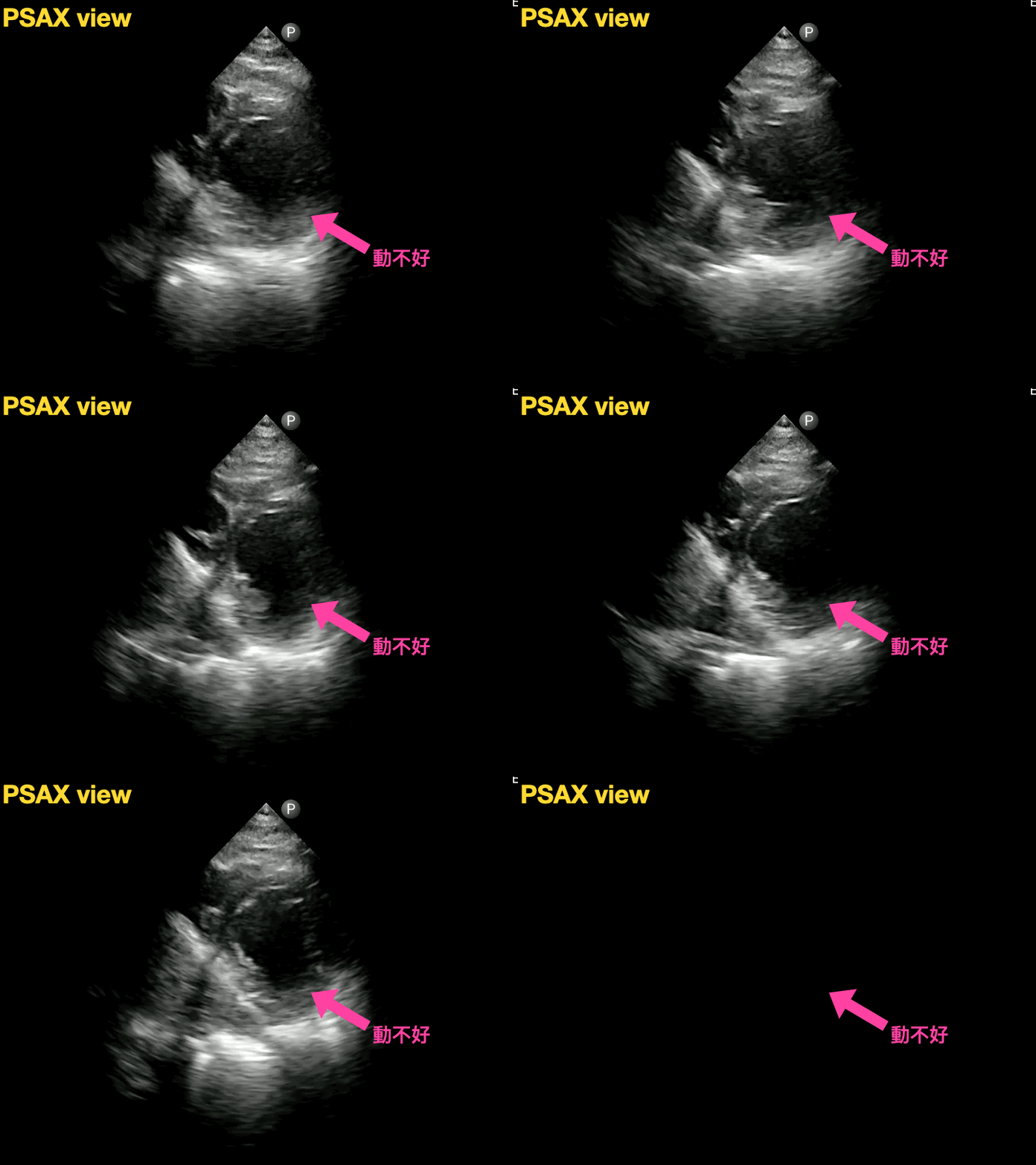

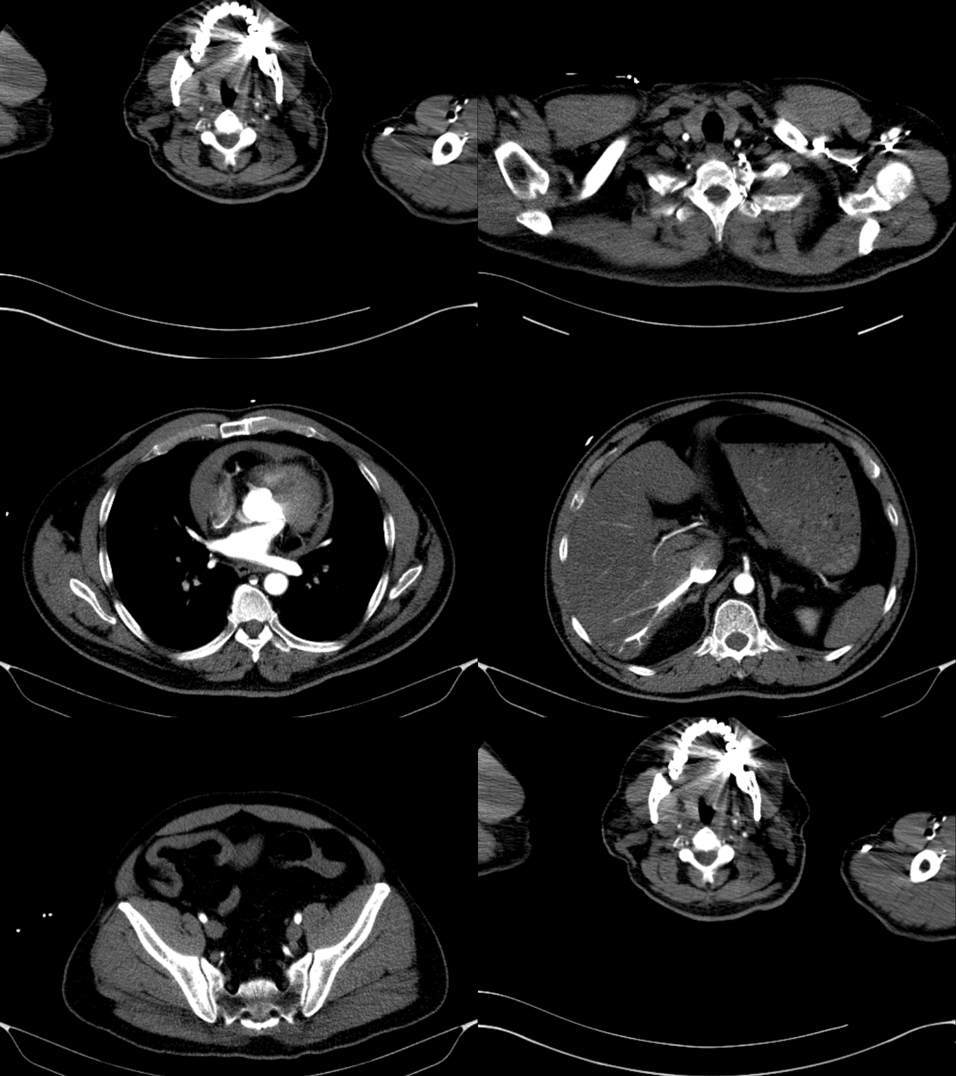
I did the heart POCUS after the patient came back from the CT room to the resus room, on a restless patient in crushing chest pain. I really couldn't get good views. As soon as the chest CTA showed no aortic dissection, it was time to rush to the cath lab.
What happened?
From the ECG reading, we can tell there should be an OMI (Occlusion MI): an occluded vessel. And the patient may have had a previous MI, hence the obvious QS waves in V1-V4. If you look only at the STE in V1-V4, V4 is the most worrying lead (not V2–V3). Of course, besides V4, the STE and ST segment morphology in V5-V6/I/aVL are all abnormal too. And there's reciprocal STD change in the inf.leads.
The ECG + symptoms alone should have sent him straight to the cath lab.
But......... why was there so much PEF?
Looking at the heart POCUS (Fig.5), you can see the lateral wall and apex were moving very poorly. It should be a problem with the LAD or LCx or proximal LAD.

The chest CTA (Fig.6) showed hemopericardium, with the PEF having the density of bloody fluid. And after contrast injection, the contrast couldn't be pulled back to the heart and pooled in the IVC/SVC and hepatic vein/renal vein, which tells us the patient's LV systolic function had become very poor and he was close to collapse. Possibly a double hit of MI + impending cardiac tamponade.
Free wall rupture? That was the answer I came up with
Cath report: LAD-P total occlusion

Let's look at this article4:
Mechanical Complications of Acute Myocardial Infarction: A Review. JAMA Cardiology
Mechanical complications of AMI include:
- LV free wall rupture
- Ventricular septal rupture
- Papillary muscle rupture
- Pseudoaneurysm/True aneurysm
Mechanical complications most commonly occur within 1 week after AMI. The common presentations are cardiogenic shock and acute pulmonary edema
Echo is usually the earliest tool that can pick them up. When a patient develops a mechanical complication, stabilizing hemodynamics usually takes medical Tx, and ECMO support is also needed. Surgery is the definitive Tx, but the timing of intervention is still unclear.
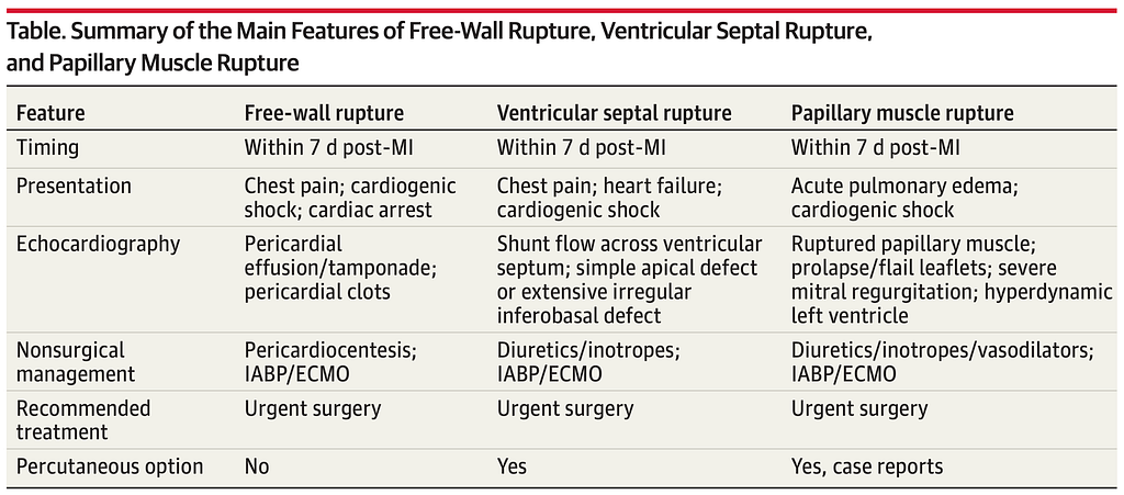
Table 1 is the table from the article; it still recommends urgent surgery for patients with free wall rupture.
Free wall rupture can be divided into three types
- type 1 occurs within 24hr
- type 2 is a slow tear at a site of localized myocardial erosion
- type 3 is rupture of a thin-walled aneurysm, usually within one week after MI
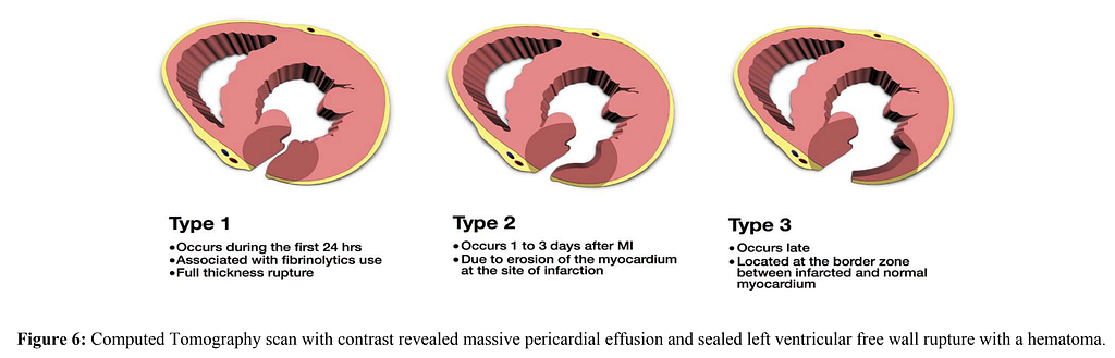
In the SHOCK trial5, there was no sex difference in free wall rupture. But patients with free wall rupture were less likely to have T2DM and old MI. The presumption is that patients with T2DM and old MI are more likely to have developed collateral circulation, which can protect the myocardium from acute rupture when a vessel suddenly occludes.
Free wall rupture within one week may also be related to straining, such as coughing or vomiting. This reminds me of the story of one of my tragic patients....... I'll write it up when I have time.
Can LVG (Left ventriculography) show the hole?
- If blood isn't spurting out, you may not be able to see where the contrast is leaking
What's the point of surgery?
- Surgery is the definitive Tx for free wall rupture; the goal is to close the tear and prevent re-rupture and pseudoaneurysm formation
Is NOM (Non-operative management) for free wall rupture very successful?6
- With NOM for free wall rupture, the survival rate may be only 10%
After free wall rupture is diagnosed → the first step of treatment is stabilizing hemodynamics (with pericardiocentesis), IABP, fluids, and vasopressors
- Because of the poor prognosis of NOM → surgery is still the first choice. Doing CAG before surgery helps the surgeon understand the state of the coronary arteries and choose a more appropriate operation.
- If NOM is chosen → stabilize hemodynamics, prolonged bed rest, strict blood pressure control, and IABP
Key takeaways:
- When any one lead in V1-V4 has a QS wave and you need to distinguish acute STEMI vs. old MI with persistent STE → you can apply the T/QRS ratio >0.36 rule
- What are the prerequisites for using the T/QRS ratio?
- T/QRS ratio >0.36 may give a false-negative when the patient's chest pain has lasted more than 6 hours
- RAD can also be seen in lateral wall MI, but overall it's uncommon in STEMI
- What proportion of each type of STEMI shows reciprocal STD change? Note especially that in ant. wall STEMI, the absence of reciprocal STD change in the inf.leads does not mean the ant. wall STEMI is fake
- What are the mechanical complications after AMI? How are they treated?
Additional references: 1 7 2 3 4 5 6
Amal Mattu’s ECG Case of the Week — December 28, 2020 — ECG Weekly — Link ↩︎ ↩︎
Smith, S. W. (2005). T/QRS ratio best distinguishes ventricular aneurysm from anterior myocardial infarction. The American Journal of Emergency Medicine, 23(3), 279–287. https://doi.org/10.1016/j.ajem.2005.01.003 ↩︎ ↩︎ ↩︎
Klein, L. R., Shroff, G. R., Beeman, W., & Smith, S. W. (2015). Electrocardiographic criteria to differentiate acute anterior ST-elevation myocardial infarction from left ventricular aneurysm. The American Journal of Emergency Medicine, 33(6), 786–790. https://doi.org/10.1016/j.ajem.2015.03.044 ↩︎ ↩︎ ↩︎
Gong, F. F., Vaitenas, I., Malaisrie, S. C., & Maganti, K. (2021). Mechanical Complications of Acute Myocardial Infarction: A Review. __JAMA Cardiology__, __6__(3), 341–349. https://doi.org/10.1001/jamacardio.2020.3690 ↩︎ ↩︎
Slater, J., Brown, R. J., Antonelli, T. A., Menon, V., Boland, J., Col, J., Dzavik, V., Greenberg, M., Menegus, M., Connery, C., & Hochman, J. S. (2000). Cardiogenic shock due to cardiac free-wall rupture or tamponade after acute myocardial infarction: a report from the SHOCK Trial Registry. __Journal of the American College of Cardiology__, __36__(3, Supplement 1), 1117–1122. https://doi.org/10.1016/S0735-1097(00)00845-7 ↩︎ ↩︎
Yan, L., Wang, H., Su, B., Fan, J., Wang, M., & Zhao, X. (2021). Survival after left ventricular free wall rupture following acute myocardial infarction by conservative treatment. __The American Journal of Emergency Medicine__, __39__, 21–23. https://doi.org/10.1016/j.ajem.2020.08.035 ↩︎ ↩︎
Dr. Smith’s ECG Blog: Chest pain and ST Elevation. — link ↩︎



Thoughts on this case? Leave a comment, a like, or a reaction below.
comments powered by Disqus