Translated from the original Traditional Chinese post. Read the Chinese original →

Diagnosing AMI in LBBB or paced rhythm: have you filled in all the gaps?
First, a question
Can LBBB be treated as AMI?
Way back when, we were taught that a new LBBB should be treated just like a STEMI: call the CV man and rush to the cath lab!!!
But study after study showed that's not the case. The proportion of these LBBBs that are true AMIs is low (leading to a high false positive rate), and patients who don't actually have an AMI end up getting PCI or rTPA, which may do them more harm than good.
So the 2013 JACC STEMI guideline¹ took the position that most LBBBs are probably old (with no old ECG to compare whether the LBBB is new), and that a new or presumably new LBBB at presentation is rare. Therefore it should not be treated as AMI.
Key point → LBBB⧧STEMI
Why is AMI hard to diagnose in LBBB or paced rhythm?
Normally, in LBBB/RBBB, in 3 important leads (I, V1, V6), the ST segment and T wave should point opposite to the direction of the terminal deflection of the QRS (secondary (2°) ST-T wave change). This is called the ST Opposition Rule
Because LBBB/RBBB both change the sequence of ventricular depolarization, the QRS morphology changes ➡ this conduction defect also changes ventricular repolarization ➡ producing secondary (2°) ST-T wave change (it's called secondary mainly because it's a change produced by the conduction defect)²
It's exactly this change that makes AMI so hard to diagnose in patients with RBBB/LBBB!!!

Not easy to understand!!!
Anyway, just remember: because the left/right bundle branch is blocked, the sequence of ventricular depolarization changes, so RBBB/LBBB has its own characteristic normal appropriate discordance (Fig 1).
If the QRS is (+) → there will be STD and TWI
If the QRS is (-) → there will be STE and an upright T wave
So when you see concordant STE or STD, be careful: has an AMI wiped out the changes a normal RBBB/LBBB should have (appropriate discordance)!!!!
Using the possible changes above:
So in 1996, Sgarbossa proposed the Sgarbossa criteria in NEJM³
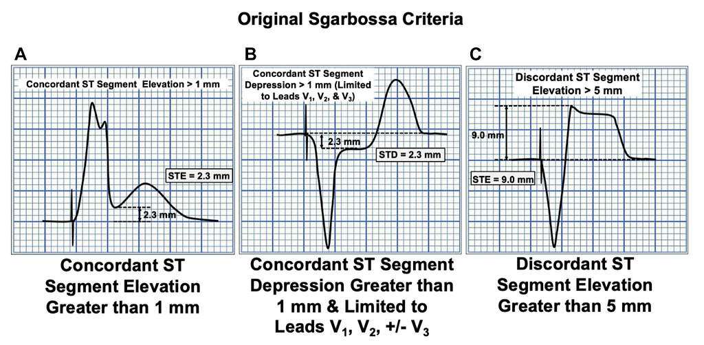
A and B are the concordant STE (A) and STD (B) described above
If A is met → 5 points
If B is met → 3 points
If C is met → 2 points
At ≥3 points, strongly consider AMI → in other words, if A or B is there, watch out!!!
Criterion C of the Sgarbossa criteria isn't strong enough. Why? Look at the figure below
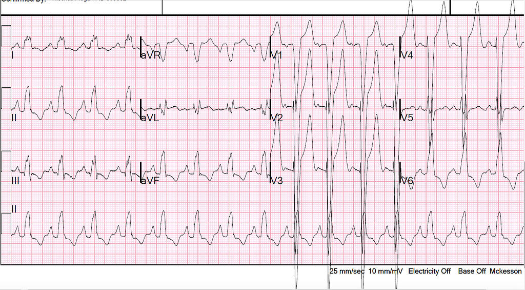
The ECG above is LBBB. It doesn't meet Sgarbossa criteria A or B, but V2~4 all meet criterion C → that is, there's discordant STE ≥5 mm . So is this an AMI? Unfortunately, in the end there was no occluded vessel.
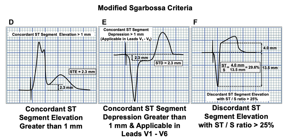
Because this finding gives false positives so easily, Smith published the Modified Sgarbossa criteria (MSC) in AEM in 2012⁴.
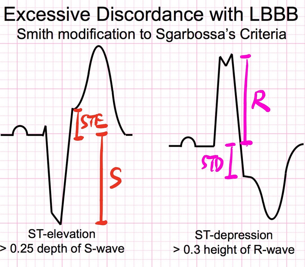
The Modified Sgarbossa criteria replaced the weak Sgarbossa criterion C with Fig 2 above. Why replace it with that? Because, as we said earlier, RBBB/LBBB has appropriate discordance, but when it's too discordant, that's not OK either.
Too much can mean two things. One is the plain measured value; the other is the proportion.
Smith found that using discordant ST elevation ≥5 mm (plain measured value) alone gives false positives easily, but switching to the proportion catches more true AMIs.
In a typical LBBB, the STE/S ratio in V1~V4 is 0.11⁴; if the STE/S ratio is >0.2, watch out for AMI⁵. The figure below (Fig 3) is from a 2015 paper in AHJ by Smith's partner Meyers, a validation study of Smith's MSC. You can see from the figure that the weighted Sgarbossa has low sensitivity but very high specificity (meaning that not seeing it doesn't mean there's no AMI, but if you do see it, you can almost diagnose AMI).
With the MSC, setting the STE/S ratio cutoff at 0.2 raises the sensitivity to 84%, while the specificity drops only a little, to 94%. With a cutoff of 0.25, sensitivity drops to 80%, but specificity can go up to 99%. With a cutoff of 0.3, you may miss many AMI patients.
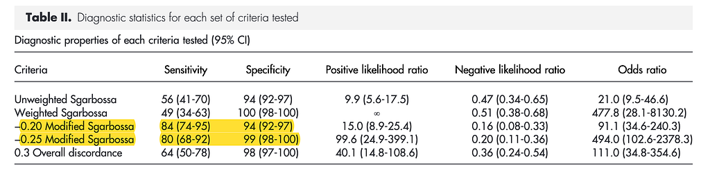
Key point → MSC rule C: STE/S ratio >0.25, think AMI (>0.2, already be careful)
Last year an AHA paper described a new way to assess for AMI in LBBB patients (the BARCELONA criteria)⁶.
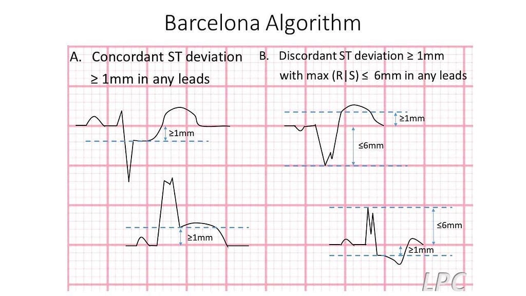
Under the BARCELONA criteria (any lead), if the patient meets either of the following two, AMI is considered possible
A. Concordant ST deviation (STE or STD) ≥ 1 mm (i.e., Sgarbossa criteria A and B, except that Sgarbossa criterion B is limited to V1~V3, while in the BARCELONA criteria any lead counts)
B. Discordant ST deviation (STE or STD) ≥ 1 mm with max (R|S) ≤6 mm (i.e., when R+S ≤6 mm, discordant ST deviation ≥ 1 mm meets the criterion)
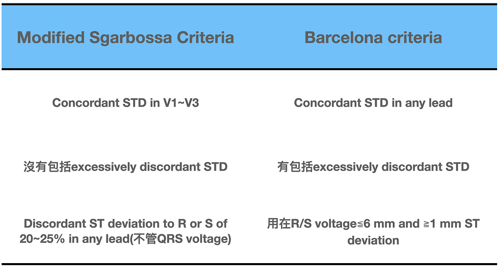
An episode of Amal Mattu ECG weekly covered the BARCELONA criteria, and the lecture included a table (Fig 5) with the comparison from this paper⁶. The first number is from the derivation cohort and the second from the validation cohort. You can see that when they validated Smith's MSC, both its sensitivity and specificity came down. Amal thinks these numbers (the sensitivity/specificity of the BARCELONA criteria) look pretty, but still need an external validation study to back them up.
Of course, Smith is no pushover either. Seeing the fruit of his hard work get knocked down, of course he had to hit back. Want to see how? Look here

Finally, the last part.
This month, on June 22, Smith's team published a paper in AEM. It's about whether the MSC can be applied to diagnose AMI in patients with a paced rhythm⁷. This is because, just like RBBB/LBBB, patients with a paced rhythm have an altered conduction pathway, so AMI is hard to diagnose. But more and more cases have shown that the MSC can actually be used in paced-rhythm patients, just as the MSC is used to diagnose AMI in LBBB.
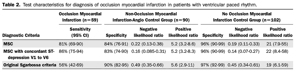
This paper used the original MSC to assess for AMI in paced-rhythm patients, but extending criterion B from V1~V3 to V1~V6 was found to raise sensitivity from 81% to 86% (Fig 7), with about the same specificity. This study shows that in patients with a pacemaker, the MSC (note: changing V1 to V3 into V1 to V6 increases sensitivity) can indeed be used to look for AMI as well.

I'll close with a flowchart (Fig 8) from the paper⁷.
When we run into LBBB/paced rhythm, the MSC can help us judge whether the patient has an AMI. If the MSC is met, consult the CV man as early as possible to evaluate for PCI and salvage the remaining myocardial cells.
- 2013 ACCF/AHA Guideline for the Management of ST-Elevation Myocardial Infarction. Journal of the American College of Cardiology, 61(4), e78–e140. https://doi.org/10.1016/j.jacc.2012.11.019
- https://www.amazon.com/ECG-2014-Pocket-Brain-Expanded-Ken-Grauer/dp/1930553250
- Sgarbossa, E. B., Underwood, D. A., & Wagner, G. S. (1996). Electrocardiographic Diagnosis of Evolving Acute Myocardial Infarction in the Presence of Left Bundle-Branch Block. The New England Journal of Medicine, 334(8), 7.
- Smith, S. W., Dodd, K. W., Henry, T. D., Dvorak, D. M., & Pearce, L. A. (2012). Diagnosis of ST-Elevation Myocardial Infarction in the Presence of Left Bundle Branch Block With the ST-Elevation to S-Wave Ratio in a Modified Sgarbossa Rule. Annals of Emergency Medicine, 60(6), 766–776. https://doi.org/10.1016/j.annemergmed.2012.07.119
- Meyers, H. P., Limkakeng, A. T., Jaffa, E. J., Patel, A., Theiling, B. J., Rezaie, S. R., Stewart, T., Zhuang, C., Pera, V. K., & Smith, S. W. (2015). Validation of the modified Sgarbossa criteria for acute coronary occlusion in the setting of left bundle branch block: A retrospective case-control study. American Heart Journal, 170(6), 1255–1264. https://doi.org/10.1016/j.ahj.2015.09.005
- Di Marco, A., Rodriguez, M., Cinca, J., Bayes‐Genis, A., Ortiz‐Perez, J. T., Ariza‐Solé, A., Sanchez‐Salado, J. C., Sionis, A., Rodriguez, J., Toledano, B., Codina, P., Solé‐González, E., Masotti, M., Gómez‐Hospital, J. A., Cequier, Á., & Anguera, I. (2020). New Electrocardiographic Algorithm for the Diagnosis of Acute Myocardial Infarction in Patients With Left Bundle Branch Block. Journal of the American Heart Association, 9(14). https://doi.org/10.1161/JAHA.119.015573
- Dodd, K. W., Zvosec, D. L., Hart, M. A., Glass, G., Bannister, L. E., Body, R. M., Boggust, B. A., Brady, W. J., Chang, A. M., Cullen, L., Gómez-Vicente, R., Huis in ‘t Veld, M. A., Karim, R. M., Meyers, H. P., Miranda, D. F., Mitchell, G. J., Reynard, C., Rice, C., Salverda, B. J., … Taylor, A. M. (2021). Electrocardiographic Diagnosis of Acute Coronary Occlusion Myocardial Infarction in Ventricular Paced Rhythm Using the Modified Sgarbossa Criteria. Annals of Emergency Medicine, S0196064421002493. https://doi.org/10.1016/j.annemergmed.2021.03.036



Thoughts on this case? Leave a comment, a like, or a reaction below.
comments powered by Disqus