Translated from the original Traditional Chinese post. Read the Chinese original →
Still Hyperkalemic?
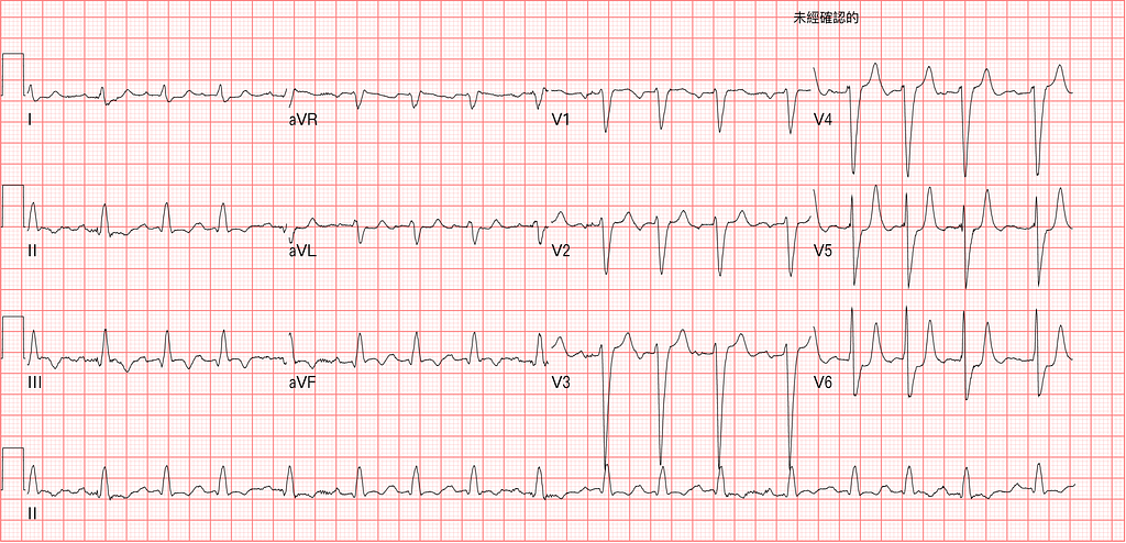
A dialysis patient came to the ED short of breath. The X-ray looked like pulmonary edema — probably fluid overloaded. Fig 1 is the ECG on arrival. Potassium: 6.1
When the patient was handed over to me, it was right when the second cardiac enzymes and potassium were due to be followed up.

Fig 2 is the ECG done at the time of the second cardiac enzyme draw.
After looking at it, I immediately ordered Calcium, D50W+RI, Kalimate, NaHCO3.
The nurse asked me: Dr. Bear, are you sure you want to give potassium-lowering meds "again"?
Dr. Bear: Yes!!!
Potassium on follow-up: 7.4
Later I looked through the chart: the previous shift had already given potassium-lowering meds: NaHCO3, Kalimate
The ECG changes from hyperkalemia are endlessly variable. Anything can show up.
It can look like the ST elevation of an MI, or like the ST depression of subendocardial ischemia. It can be slow, it can be fast.
A few key points to hold on to:
- If ST elevation shows up in the Rightward leads (aVR, V1, V2, III), think Hyper-K, especially with RAD on top (also put it in the DDx alongside PE and Na channel blockers)
- If you see Narrow based Peaked T waves, also think Hyper-K (like the insanely pointy T waves in V3~5 in Fig 2) → note that the sensitivity of Peaked T waves isn't high (meaning: no Peaked T waves doesn't mean no Hyper-K)
- A slow heart rate with a conduction block should also make you think of it (P waves flattening)
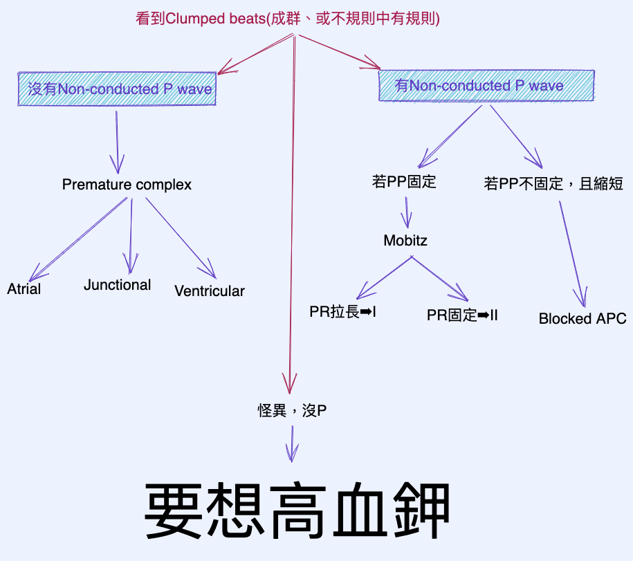
- If you see Clumped beats, and they look bizarre with no P, think of it too (see the flowchart in Fig 3)



Thoughts on this case? Leave a comment, a like, or a reaction below.
comments powered by Disqus