Translated from the original Traditional Chinese post. Read the Chinese original →
Can You Tell It's an MI?
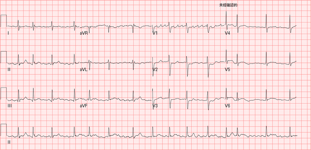
A grandma in her 70s. Chest pain started in the afternoon, bad enough that she broke into a cold sweat. She got to the hospital 2~3 hours later.
Usually V1 and II are where you can see P waves most clearly, i.e., what the atrium is up to.
In V1 there seem to be a lot of P waves.
If it's AFL, the atrial rate is about 250~350 bpm. If it's Af, the atrial rate is about 350~600 bpm.
If these were flutter waves, the usual inverted P waves don't seem to show up in the inf. leads.
It should be Af. In V1, some of the PP intervals are only 3~4 small boxes, over 350 bpm. (Fig.1)

When I read an ECG, I usually go in this order: Rate, Rhythm, Axis, interval, ischemia, to see whether anything urgent is going on.
Rate: look at the atrial rate and the ventricular rate, i.e., what the atria and the ventricles are doing.
Atrial rate: (look at the P waves)
- 100~250 bpm: atrial tachycardia
- 250~350 bpm: atrial flutter
- 350~600 bpm: atrial fibrillation
Ventricular rate: (look at the QRS; based on the ventricular rate, figure out where the firing is coming from)
- 20~40 bpm: Purkinje fibers firing
- 40~60 bpm: AV node/His bundle firing
- 60~100 bpm: SA node firing
Rhythm: tells us whether the rhythm is regular or irregular.
You can mark an RR interval with a line on a piece of paper and compare it with the next RR interval. Of course, with calipers you don't have to waste paper.
EKG Caliper Black ECG Calipers Electrocardiogram Divider for Nursing
That's the Amazon link up there. I personally find these really handy. Except at work people occasionally ask me why on earth I'm carrying a drawing compass around XD (not sponsored)
Once you've sorted regular vs. irregular, of course there's a whole set of DDx that comes up.
Axis: pay special attention to RAD

Different axes can come with different DDx lists. (Fig.2)
I'm an EM doc, so I'm a bit more cautious when RAD shows up.
Here are a few golden lines about RAD that the masters often say.
1️⃣ Watch out, as Amal Mattu often says: RAD + STE in the rightward leads, think especially about PE, Na channel blockers, Hyper-K
- Which leads are the rightward leads? ➡︎ aVR/V1 and/or V2/III
2️⃣ AMI rarely presents with RAD and tachycardia. If you see AMI + tachycardia, it's usually already cardiogenic shock
Interval: look at the PR interval, QRS, and QT interval
With the QT interval, watch for too long or too short? Each brings different problems!
The QT interval has to be corrected for rate.
If QTc>500 ms, be careful. A prolonged QT always has a reason behind it.
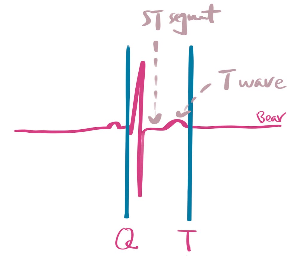
From Fig.3 you can see that a prolonged QT interval can come from a longer ST segment or a bigger T wave (T-U fusion, which looks like a bigger T wave, or a genuinely bigger T wave -> Hyperacute T wave)
- Longer ST segment: commonly Hypo-Ca and hypothermia
- Bigger T wave: commonly Hypo-K, Hypo-Mg, IICP, AMI, Na channel blockers
So what actually happens with QT prolongation? Why do we get so nervous about it?

Fig.4 is the QT interval nomogram1. Above this line, there's a risk of TdP.
A few key points to understand about this figure. (Note that the figure uses the QT interval)
- H.R<60 bpm: using the 1/2-RR-interval rule of thumb for QT prolongation can be risky. In other words, with H.R<60, just take 485 ms as the cutoff point to watch for TdP; don't use 1/2 the RR interval
- H.R>60 bpm: at different H.R, the QT interval at which TdP occurs is different
- H.R 100 bpm: take about 400 ms to gauge TdP risk
So what conditions come with a shortened QT?
In the latest ECG Weekly2, Amal Mattu covers exactly this question.
Let me highlight the key points.
Men: QTc ≤ 330 ms
Women: QTc ≤ 340 ms
Or if you see a QT interval < 400 ms → think short QT interval
Common causes include:
- Digoxin toxicity
- Hyper-Ca
- Hypothermia
- Short QT syndrome (SQTS)
Finally………we get to ischemia……Orz (that tangent went way too far)
Back to the main topic: did this grandma actually have an MI (AMI)?
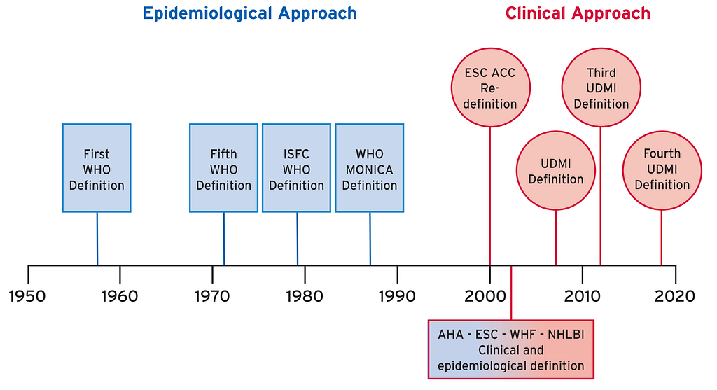
In the 1950s~1970s, the WHO defined MI based on ECG changes. Later biomarkers were added, and in 2000 the ESC/AHA began redefining MI.
Let's first look at the Fourth Universal Definition of MI published in 2018 by the ESC/ACC/AHA/WHF. I roughly translated the original definition into Chinese so it's easier to follow.
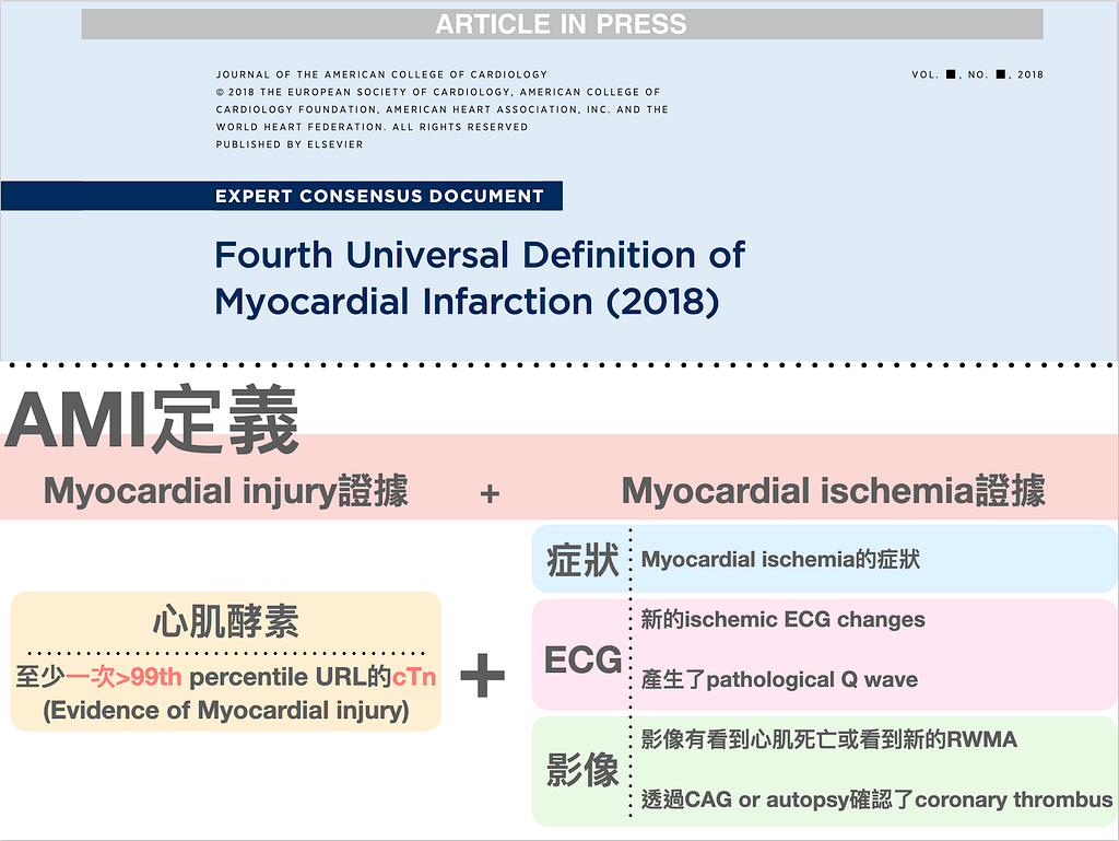
AMI = evidence of myocardial injury + evidence of myocardial ischemia
- What's the evidence of myocardial injury → a biomarker value >99th percentile at least once (the heart got hurt)
- What's the evidence of myocardial ischemia? → basically nothing more than symptoms / ECG changes / imaging findings
Let's look at this patient's POCUS images.
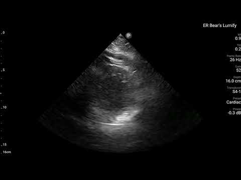
The patient was solidly built, so image acquisition wasn't easy, but you can faintly see the Ant./Lateral/Inf walls all moving poorly, while the septum still contracts pretty well!
The patient's subsequent TnI was above the 99th percentile of URL.
So did this patient have an MI → yes (because there was both evidence of myocardial injury and evidence of ischemia)
But to say for sure the patient had an AMI, by definition you still have to wait for the biomarkers. Only when it's truly above the 99th percentile of URL, or the biomarker shows a rise and/or fall, can you call it an MI.
But at the very start of an MI, biomarkers don't necessarily rise! (Fig.6)

Let's see what useful information we can get from Fig.63.
The figure shows that after an AMI, it takes a while to reach >99th percentile of URL, or for two biomarker draws to show a certain delta change (20%). In other words, very early in an AMI, the biomarkers can't rise yet.
Since biomarkers can't rise that early, it's no surprise that when an ACS patient arrives and gets the first blood draw, the TnI or TnT is negative. Never assume a negative biomarker means it's not an MI.
The ECG master Dr. Smith has said many times on his ECG Blog that, whether STEMI or OMI (Occlusion MI), the initial troponin is not a reliable indicator for diagnosing MI4.
Let's ask a question here. When a patient first arrives in the ED, how accurate are the initial STEMI criteria and the initial troponin for diagnosing OMI (Occlusion MI)?
First, the ECG:
- In a prospective validation study, applying STEMI criteria to the first ED ECG was only 21% sensitive for OMI, and disproportionately missed Inf.Post OMI5
- This paper also reported the sensitivity of STEMI criteria for AMI. The study enrolled 2486 patients presenting to the ED with chest pain (all with normal cardiac conduction). Of these, 438 had confirmed AMI, and 81 met STEMI criteria. The sensitivity was only 35%5 → so using STEMI criteria to catch patients with an occluded vessel really isn't very sensitive. In other words, many more patients with an occluded vessel don't present with a STEMI-pattern ECG.
- Another study included 41000 patients; 11% initially had a non-diagnostic ECG ➡ 1/3 converted to STEMI within 30 minutes, 50% within 45 minutes, and 75% within 90 minutes6 → in symptomatic patients, serial F/U ECGs are extremely important
Next, troponin
- Stephen Smith reported that in his study, among 925 STEMI patients, 14.4% had an initial troponin below the 99th percentile of URL7
- This study also showed that early in STEMI, 25% of true STEMI patients had hsTnI<99th percentile of URL
And because troponin isn't a reliable indicator early in MI, Dr. Smith says: if the initial ECG has OMI findings, believe it!!!!!
Case continue
Back to grandma.
Initially V2~V3 had STD, and V5~V6 had straight minimal STE.

When you see maximal STD in V1~V4, what should you think of?
Yes!!! Post.wall MI~~~~ (check out this post of mine from the end of last year)
Seeing that ECG, I felt like I'd been zapped!!!! Here we go again. Could it be another isolated post.wall MI? That's what I told myself. And even if it wasn't an isolated post.wall MI, the inf. wall/lateral wall had problems too. Minimal straight STE in the inf. leads and lateral leads → there really is a problem!!!
Use this initial ECG and STEMI criteria to convince the CV man to activate the cath lab? Seems impossible, because it doesn't meet STEMI criteria. So what about the STD in V2~V3?
Smith's post says maximal STD in V1~V4 has 96% specificity for Post.OMI. So how much STD does it take to count?
Any STD in V1~V4 has to be considered posterior OMI until proven otherwise8!!!!!
I know these findings won't get anyone to buy in (cath lab activation).
I chose to repeat the ECG in 30 minutes, and in the meantime I had time to do a cardiac echo to look for RWMA, so I'd have enough clearer evidence to convince people.
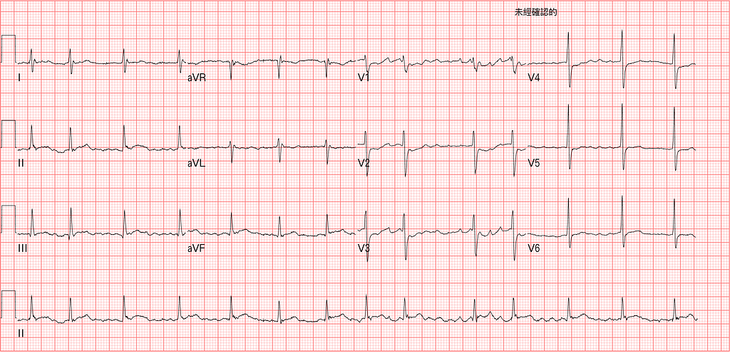
Fig.7 is the ECG 30 minutes later (symptoms ongoing). Still STD in V1~V5 (more obvious in V1), and the inf. leads still have minimal STE.
Putting together the ECG findings on both tracings and the bedside echo showing RWMA at the lateral/inf. wall:
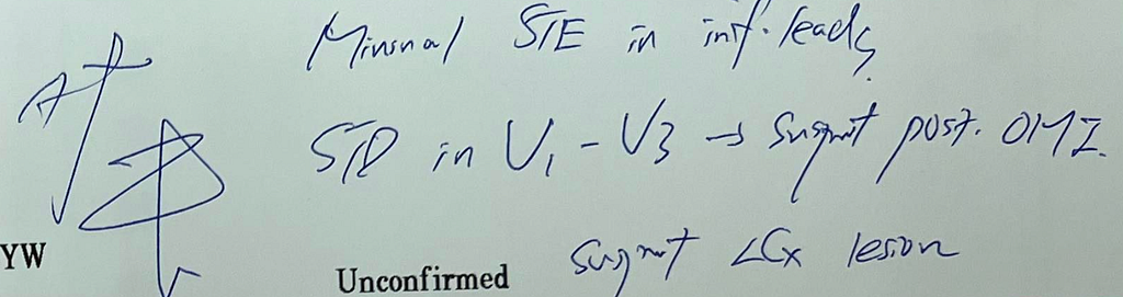
I wrote my findings on the second ECG: suspect an LCx lesion.
But the CV man thought the ECG was so-so and wanted to wait for the biomarkers.
Here's a question, then? Namely, how reliable are biomarkers for diagnosing early MI?! (Covered above, scroll back up)
If the biomarker is positive → it really does fit the MI findings on ECG + echo
If the biomarker is negative → it can't rule out an occluded vessel
After the second ECG, the patient's symptoms eased for a short while. Then they got worse again (probably re-occlusion). I called CV again, and CV still wanted to admit the patient to the ICU first and do PCI the next day.
I have to bring up again a figure from the 2020 ESC NSTEMI Guideline that I've mentioned many times on the blog. (Fig.8)
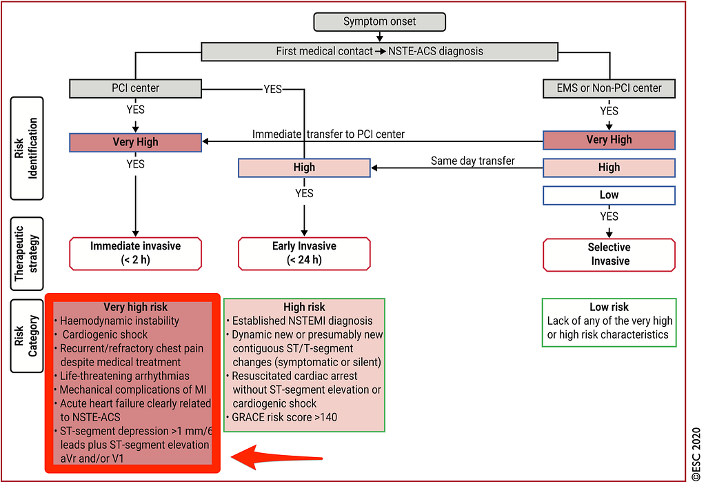
Why bring up this figure? This patient didn't meet STEMI criteria but definitely had an MI, so under the current guideline, she falls under NSTE-ACS. The very high risk group in the lower left corner should get immediate invasive (PCI) within <2 hr. The patient's symptoms were recurrent and persistent, so she belonged in this group.
The patient had PCI the next day: LCx total occlusion

Key takeaways:
- When reading an ECG, go through the basics, Rate, Rhythm, Axis, interval, ischemia, so you're less likely to miss important clues
- For rate, look at both the atrial rate and the ventricular rate
- For axis, watch for RAD
- What problems come with QT prolongation and QT shortening?
- What's the definition of AMI?
- How much do the initial ECG and troponin help in diagnosing MI?
- Maximal STD in V1~V4 → highly suspect Post.OMI until proven otherwise (any STD has to be considered)
- What are the criteria for immediate invasive in NSTE-ACS?
Additional references: 2 1 3 4 5 6 7 8
Chan, A., Isbister, G. K., Kirkpatrick, C. M. J., & Dufful, S. B. (2007). Drug-induced QT prolongation and torsades de pointes: evaluation of a QT nomogram. __QJM: Monthly Journal of the Association of Physicians__, __100__(10), 609–615. https://doi.org/10.1093/qjmed/hcm072 ↩︎ ↩︎
Amal Mattu’s ECG Case of the Week — February 14, 2022 — ECG Weekly — link ↩︎ ↩︎
Thygesen, K., Alpert, J. S., Jaffe, A. S., Chaitman, B. R., Bax, J. J., Morrow, D. A., & White, H. D. (2018). Fourth Universal Definition of Myocardial Infarction (2018). __Journal of the American College of Cardiology__, __72__(18), 2231–2264. https://doi.org/10.1016/j.jacc.2018.08.1038 ↩︎ ↩︎
Dr. Smith’s ECG Blog: Which ACS had more myocardial damage? The one that meets STEMI criteria, or the one with the ‘normal’ ECG? — link ↩︎ ↩︎
Hillinger, P., Strebel, I., Abächerli, R., Twerenbold, R., Wildi, K., Bernhard, D., Nestelberger, T., Boeddinghaus, J., Badertscher, P., Wussler, D., Koechlin, L., Zimmermann, T., Puelacher, C., Rubini Gimenez, M., du Fay de Lavallaz, J., Walter, J., Geigy, N., Keller, D. I., Reichlin, T., & Mueller, C. (2019). Prospective validation of current quantitative electrocardiographic criteria for ST-elevation myocardial infarction. __International Journal of Cardiology__, __292__, 1–12. https://doi.org/10/gnkvjh ↩︎ ↩︎ ↩︎
Riley, R. F., Newby, L. K., Don, C. W., Roe, M. T., Holmes, D. N., Gandhi, S. K., Kutcher, M. A., & Herrington, D. M. (2013). Diagnostic time course, treatment, and in-hospital outcomes for patients with ST-segment elevation myocardial infarction presenting with nondiagnostic initial electrocardiogram: A report from the American Heart Association Mission: Lifeline program. __American Heart Journal__, __165__(1), 50–56. https://doi.org/10.1016/j.ahj.2012.10.027 ↩︎ ↩︎
Wereski, R., Chapman, A. R., Lee, K. K., Smith, S. W., Lowe, D. J., Gray, A., & Mills, N. L. (2020). High-Sensitivity Cardiac Troponin Concentrations at Presentation in Patients With ST-Segment Elevation Myocardial Infarction. __JAMA Cardiology__, __5__(11), 1302. https://doi.org/10.1001/jamacardio.2020.2867 ↩︎ ↩︎
Dr. Smith’s ECG Blog: Any ST depression in V2 and V3 is posterior OMI until proven otherwise, especially if downsloping — link ↩︎ ↩︎



Thoughts on this case? Leave a comment, a like, or a reaction below.
comments powered by Disqus