Translated from the original Traditional Chinese post. Read the Chinese original →
Arriving hypotensive and hypoxic: is there something wrong with the ECG?
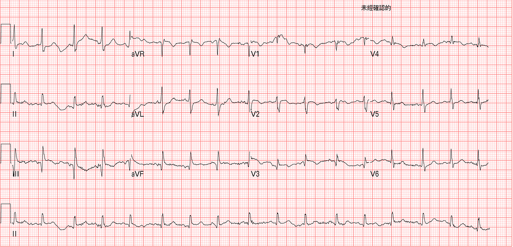
72-year-old woman, brought to our hospital by ambulance, wee-woo~~~~wee-woo.
That day the ED was, as usual, boarding hell: patients who couldn't get an inpatient bed. After handover from the night-shift doc at 07:30, I already had a whole string of patients on my hands.
About an hour into the shift, triage wheels a patient in from outside.
Parks her right in front of my desk.
Dr. Bear in Room 2, your patient: hypoxic, hypotensive.
Hearing she was mine, I hurried over to see her.
She was on the NRM at 15 L/min the EMTs had put on, SaO2 about 96%. The EMTs said that in the ambulance her SaO2 was around 40–50%.
SBP close to 90, poor SaO2, breathing fast, altered. She looked like she already had her left foot through the door of the King of Hell's place.
I immediately explained to the family that she was critically ill: things were very bad, and she could take a life-threatening turn at any moment.
The ECG got done fast. Let's read it~~
Rate:99 bpm
Rhythm:SR
Axis:Normal axis
Interval:No QT prolong
Ischemia:Q in III/aVF, TWI in V1–4, III
First, ask yourself a few questions:
- In which leads is a Q wave normal? What is a pathologic Q wave?
- Is the S wave in lead I normal?
- How do you see that V1-V4 in Fig.1 have TWI?
- What could the TWI in V1-V4 be?
In which leads is a Q wave normal? What is a pathologic Q wave?
First, take a look at a figure from ECG master Ken Grauer (Fig.2)
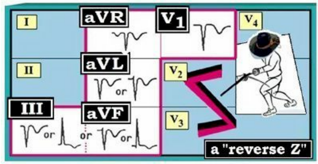
- This figure is the reverse Z shape mnemonic. That is, if you find a Q wave and/or TWI in just these five leads (III/aVF/aVL/aVR/V1) on their own, it's very likely normal. (Caveat: young person, no heart problems)
- In some leads, even a large Q wave isn't necessarily infarction → these leads include III/aVL/aVR/V1
- Septal q waves can show up in one or more lateral leads (I/aVL/V4/V5/V6) → these q waves should not be confused with pathologic Q waves
- When the Q wave is bigger and wider → the infarction has very likely been there for a while, especially if neighboring leads show the same thing, which points even more to an old one
Putting it together: the leads in the reverse Z shape + the leads where septal q waves can appear → all of these can have normal Q waves.

So how do pathologic Q waves and septal q waves differ by definition? (Fig.3)
- The textbooks say a pathologic Q wave is generally wider (>30 ms in duration), and the Q wave amplitude is > 25% of the R wave of the QRS.
- In practice we usually take 1 small box (0.04 s) as the width for a pathologic Q wave. The LITFL article also says that if it's > 2 mm deep, and if it's seen in V1-V3, you should consider the Q wave abnormal (pathologic Q wave)1
In this case, the Q waves in III/aVF are clearly abnormal.
Next: is the S wave in lead I normal?
On a normal 12 lead ECG you generally see only a small S wave in lead I, or no S wave at all. But if there's a big S wave (heads up → this is an abnormal ECG finding), you have to consider possible RV strain2.
In the DDx of axis deviation (Fig.4), we're very used to letting the presence of RAD (S wave > R wave in lead I) prompt us to think about possible pulmonary embolism
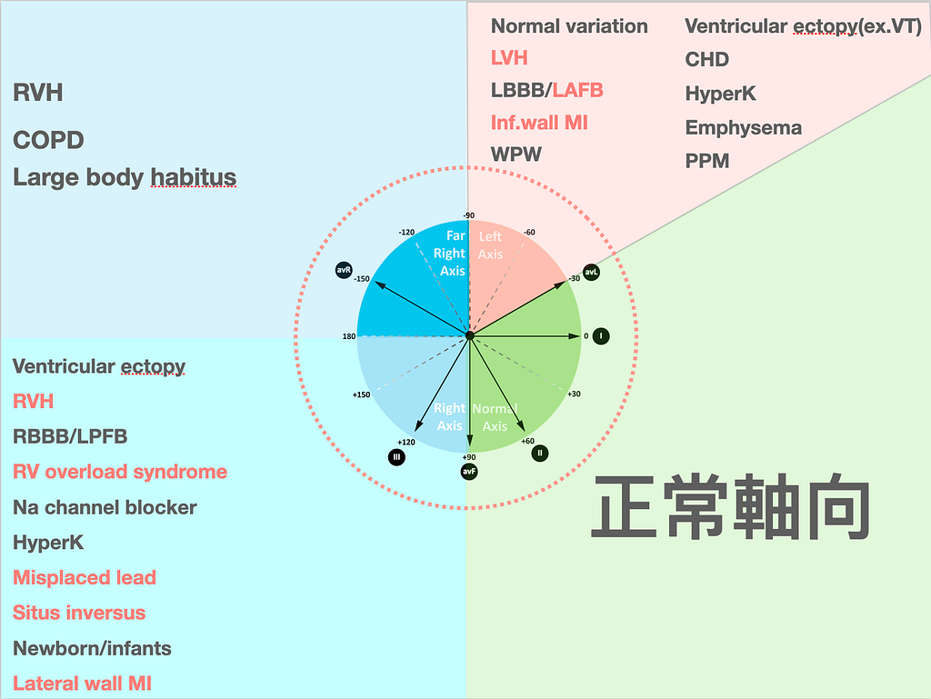
But the skill is in the details of the ECG read: be very, very careful with a big S wave. Don't rule PE out just because you don't see RAD.
ECG Tips: Watch out, a big S wave may be caused by RV strain
How do you see that V1-V4 in Fig.1 have TWI?
Why could I write down TWI in V1–4 right after looking at the arrival ECG (Fig.5)?
Once you pick up TWI in the precordial leads, in this battle with the King of Hell over who gets to take the patient, we're almost one more step ahead and able to talk back to him.
I think this reading technique is really important. Whether it's applied here or elsewhere: for example, sometimes in an RBBB patient it isn't easy to tell whether there's STE.
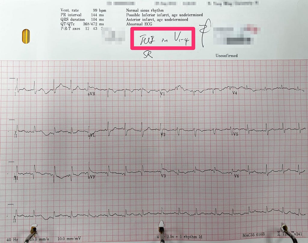
In Fig.5 the TWI in the precordial leads isn't very obvious, e.g., V2–3. That's mainly because the baseline isn't that steady (it wanders), and the downward deflection of the TWI is small, so it's hard to tell there's TWI.
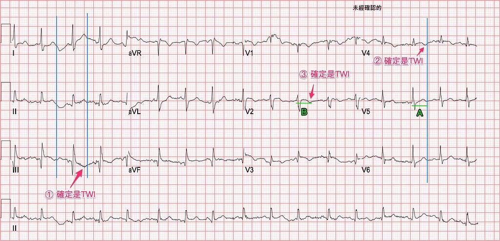
I've marked up the original ECG. Follow my explanation and let's crack it step by step: is there TWI or not?
① First, is there TWI in lead III?
- First look at the two vertical blue lines I drew; they're where I put the possible end of the T wave. Because the T wave in lead I is very clear, a vertical line dropped down from lead I has to land on the end of the T wave in leads II/III. Remember, this is the simultaneity of the 12 lead ECG along the vertical axis. You can't draw a vertical line at the end of the T wave in lead I and have it turn into, say, the start of the ST segment by the time it reaches lead II.
- So you can say with confidence that the slight dip in lead III, lined up against lead I, really is TWI.
② So is there TWI in V4?
- Using the same method, I again find that the T waves in V5–6 are very obvious. I put a vertical blue line at the end of the T waves in V5–6, and that way you can see that the tiny dip in V4 is also TWI.
③ So is there TWI in V2?
- Here we hit a hard spot: V1–3 don't have an obvious upright T wave like the other leads that we can drop a vertical line from.
- This is where another technique comes in: in a regular rhythm, the intervals in theory don't keep changing. For example, in Fig.6 horizontal green line A is the QT interval I drew (start of the QRS to the end of the T wave). So all we have to do is find the lead on the 12 lead ECG where the QT interval is clearest and most obvious, draw it out horizontally, then drag it over (e.g., green line B in this case) to the spot where we suspect possible TWI and compare. In this case that tells you clearly that the small dip in V2 is also TWI. This is the consistency of the 12 lead ECG in a regular rhythm.
ECG Tips: Make good use of the simultaneity of the 12 lead ECG (vertical lines) and the consistency of a regular rhythm (horizontal lines); this often solves a lot of hard problems
To do a good job, you first need good tools. I strongly recommend a small steel ruler and a Caliper as must-haves on shift (I'm not doing a livestream sales pitch here XD)
Feel free to buy the fine little steel ruler that comes with that Japanese-brand mystique →link
Feel free to buy the Caliper from Amazon →link
What could the TWI in V1-V4 be?
First, let's reframe the question. What it's really asking is: when TWI shows up in the R’t precordial leads, what conditions can that be?
In Amal mattu's ECG teaching, the recent 4/113 and 6/274 sessions covered which conditions you need in the DDx when TWI appears in V1–3.
Here's the DDx, straight up: (DDx of TWI in the precordial leads)
- ARVC
- RBBB
- RV strain →must be ruled out immediately
- BTWI
- Wellens’ syndrome →must be ruled out immediately
- Brugada syndrome →must be ruled out immediately
- Persistent juvenile TWI
In our ED role, when we see TWI in the R’t precordial leads, we have to immediately rule out three potentially lethal problems.
Is the TWI caused by RV strain?
Is the TWI caused by Wellens’ syndrome?
Is it Brugada syndrome?
Given this patient's symptoms and the ECG morphology, it doesn't look like Brugada syndrome, so that can be ruled out right away.
With TWI in the R’t precordial leads, when you're ruling out the lethal problems, it always ends up coming down to RV strain vs. Wellens’s syndrome, i.e., the PE vs. ACS showdown~~~
It's like how Tai Tzu-ying (Taiwan's badminton star), deep into a tournament, always ends up facing Chen Yufei……Orz (lost again, don't worry, go again!!!)
First, for the PE vs. ACS showdown, we have to understand why TWI shows up in each in the first place. Only once we understand that can we bring in the patient's history and current symptoms and go head to head.
Let's look at Wellens’ syndrome first. Wellens’s syndrome comes about because there's a brief period of vessel occlusion, but it very quickly undergoes spontaneous reperfusion on its own before a large infarction has formed. This reperfusion is what produces the Wellens’ wave on the ECG.
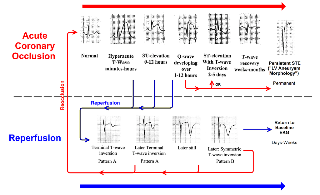
Fig.7 is from the teaching images on Smith's ECG Blog.
The top half of the figure shows the ECG changes of ACO (acute coronary occlusion), and the bottom half shows the ECG changes you'll see if the vessel opens on its own (spontaneous reperfusion) before a large infarction has formed: that is, reperfusion T waves.
These changes (meaning reperfusion) start with terminal TWI (i.e., biphasic TWI,
Pattern A): the T wave goes up first, then down. As reperfusion continues, terminal TWI then turns into deeply TWI (Pattern B).
Clinically we see deeply TWI more often (about 75%). Why do we see terminal TWI less often (about 25%)?
- Because these reperfusion T-wave changes always go from terminal TWI to deeply TWI, and the terminal TWI phase may be fairly short; if we didn't get an ECG at that moment, we just didn't see it.
As for the RV strain caused by PE, it's mainly because the pulmonary vessels are occluded, leading to RV dysfunction from RV hypertrophy or dilation. At its core, the RV heart muscle is deformed, and that's what produces the dysfunction.
So why does TWI show up in the precordial leads? This is thought to be ischemia from the low cardiac output that RV dilation and RV strain cause, but the true pathophysiology isn't fully understood yet. Take a look at this article if you want5.
Which ECG features may indicate acute pulmonary embolism?
Now here's the real point of the whole post6 (please tell me I didn't just type a whole post of filler XD)
➡️Sinus tachycardia →this isn't required to diagnose PE, but a relatively fast heart rate is a common finding (usually we use >90 bpm); in significant PE you'd expect to usually see it
➡️S1Q3T3 →not sensitive or specific for PE; it's rare for S1Q3T3 alone to diagnose a new significant PE. In other words, this ECG sign is very helpful when it shows up together with other ECG evidence for PE (it does more to support the diagnosis of PE)
- Just how low is the sensitivity of S1Q3T3? (table data7)
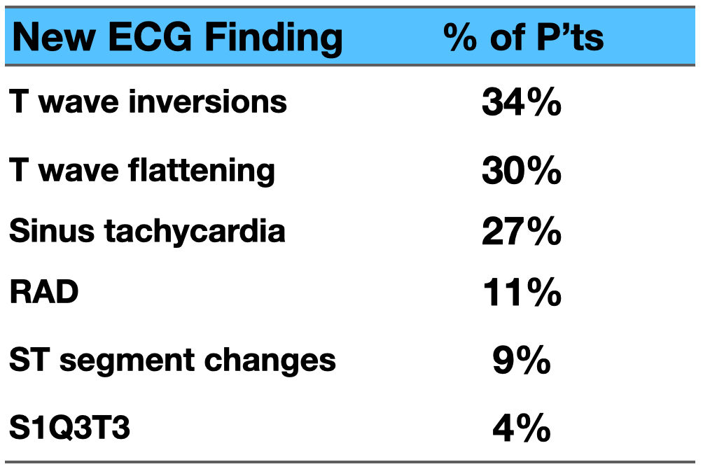
The sensitivity of S1Q3T3 is only 4%…….uh, don't count on seeing S1Q3T3 to diagnose PE. Only if you're cursed with crap luck and see 1000 PE cases (then nobody will want to work a shift with you) will you have enough S1Q3T3s to brag about.
- So according to Fig.8, what are the ECG findings that commonly show up in PE? →Add up the first and second items: that's T-wave changes (inverted or flattened), about 60%
ECG Tips: Among ECG findings in PE, T-wave changes are the most common, more common than S1Q3T3
Also, this LITFL article8 says a few things about S1Q3T3; I've written them down so we can go through them together.
The article says → there's a lack of supporting literature showing S1Q3T3 is sensitive and specific for PE, but its individual components can give us some information toward diagnosing PE
- E.g., a bigger S wave in lead I suggests clockwise rotation or RAD (suggesting the axis is shifted to the right)
- TWI in lead III can be a normal finding, but if clinically there's also right precordial leads TWI, PE should be strongly considered
- A Q wave in lead III may also be a normal variant, or may reflect regional RV ischemia or infarction (secondary to acute pressure overload)
I want to say more about point two here. A 2007 study in The American Journal of Cardiology showed that when there's R’t precordial leads TWI and the diagnosis comes down to just ACS vs. PE, TWI in lead III should make you strongly suspect PE →present in 88% of PE, but only 1% of ACS9
ECG Tips: R’t precordial leads TWI + lead III TWI → PE absolutely goes at the top of the differential
➡️RV strain present
- When there's RV strain, be careful, because it's one of the key ECG markers that a significant PE is affecting hemodynamics →in the R’t precordial leads (STD and/or TWI in V1~V3 ± inf.leads)
- This finding is very easily mistaken for coronary ischemia; be very careful
➡️RAA (Right Atrial Abnormality) present
- Definition: tall ≧ 2.5 mm, peaked and pointed P wave in inf.leads (II/III/aVF) and/or pointed P wave in V1, V2
- Medically, the situation of enlarged RA w/o RV enlargement ➡tricuspid stenosis
➡️Abnormal axis direction➡including RAD or indeterminate axis
- RAD is very common in acute PE
- An indeterminate axis can carry diagnostic value similar to RAD (meaning: in lead I the S wave is on the bigger side; it doesn't have to be like RAD, where the S wave must be clearly > the R wave ➡if the S wave is on the bigger side, consider possible PE)
- Quick refresher on what an indeterminate axis is →when the QRS complex is nearly isoelectric in all 6 limb leads, the axis can be called indeterminate
➡️PRWP
- Deep S waves in V5, V6 suggest a significant force pulling to the right ➡this article says a deep S wave in I and/or V6 suggests there may be a disease affecting the pulmonary circulation or the LV (a force pulling to the right)10
➡️aVR STE
- aVR is a rightward lead; with rightward leads STE + RAD, consider PE
- Which are the rightward leads? aVR/V1±V2/III
ECG Tips: Amal mattu used to say all the time that when you see rightward leads STE + RAD you have to think of three DDx: PE, Na channel blocker, Hyper-K
Case course:
Seeing V1–4 TWI + S1Q3T3 on her ECG, rate close to 99 bpm, the first thing that popped into my head was: could this be from RV strain?
If these ECG findings were from RV strain, and the patient was also in shock and short of breath, what tools did we have to help with the differential? She looked like the King of Hell was about to drag her off~~~


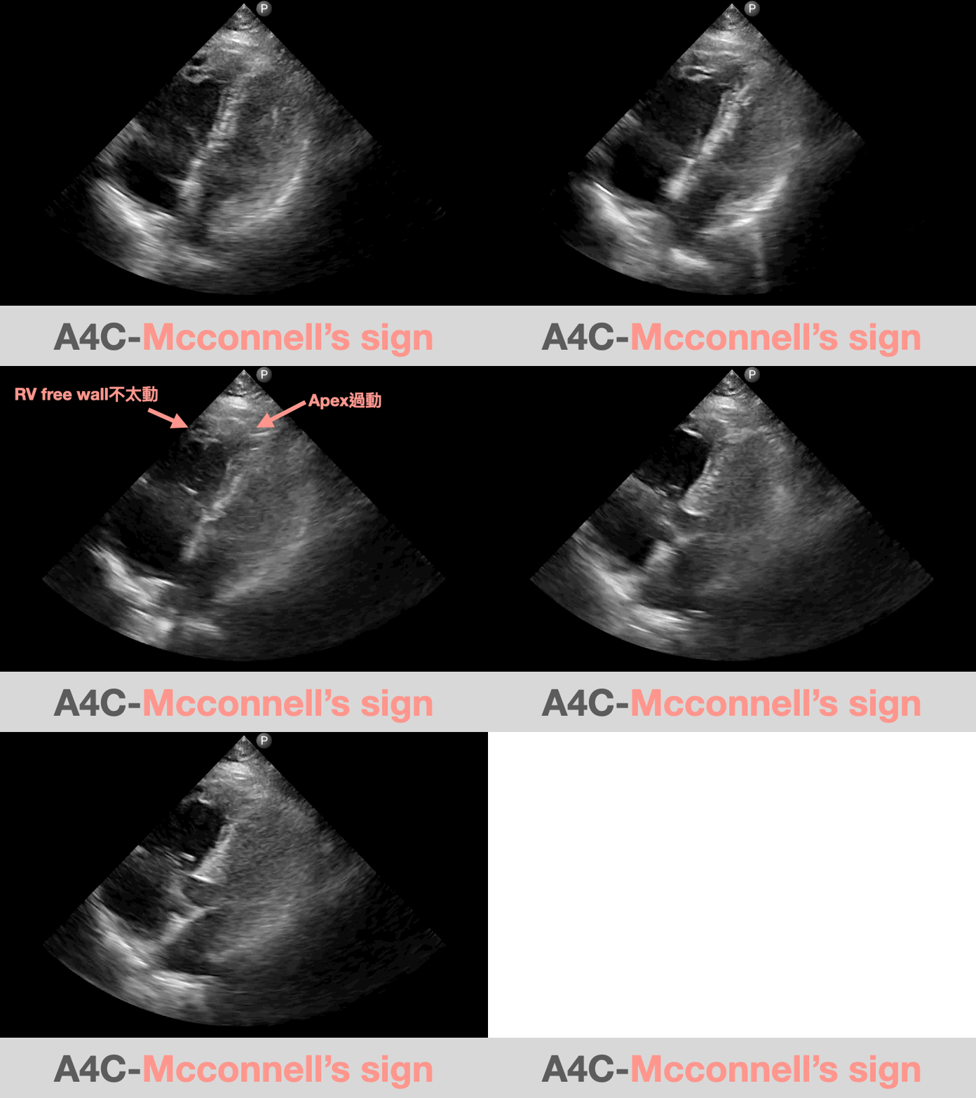
The moment I saw an obvious RV strain pattern on the ECG, I put my pricey Lumify right on her chest. After the ultrasound, I already had a clear answer in my head.
Skip the X-ray. I pushed her straight to the CT room for a chest CTA.

You can see the coronal view of the chest CTA here
The chest CTA showed both pulmonary arterial trees packed full of clot…..Orz
The chest CTA was done within about 30 minutes of her arrival.
How should this patient be treated?
As a doctor, there's one situation I really dread: you make the diagnosis, but there's nothing you can do.
Like a surgeon who pulls off the operation, but the patient dies on the table.
ED docs end up in this awkward spot a fair number of times.
A trauma patient, diagnosed with massive intra-abdominal bleeding, but there's no time to operate; the patient dies in the ED.
An aortic dissection gets diagnosed, there's no time to get to the OR, and the patient dies in the ED.
A STEMI patient collapses in the ED before going up to the cath lab, you shock and shock and can't get them back, and they die in the ED.
There are so many situations in the ED where a patient can die in our hands.
So how do we give this patient a chance at being snatched back from the King of Hell? The diagnosis is made, but is she going to die in my hands?
I had the nurse go get rTPA right away. We were going to give rTPA to dissolve the clot. I clearly remember her son pulling me aside and telling me that if there was a more expensive, better drug, just use it. (Uh….. this is the best drug NHI (Taiwan's National Health Insurance) covers.)
Remember which emergencies rTPA can be used for?
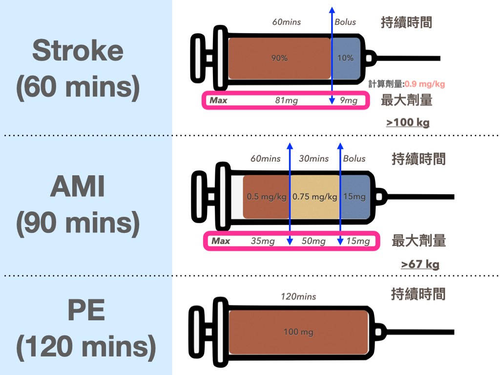
What are the key points in treating the crashing pulmonary embolism patient? (The points below come from this article11)
To save a PE patient, you first need to understand what the PE death spiral is. Before the key points, take a look at a figure (Fig.11)
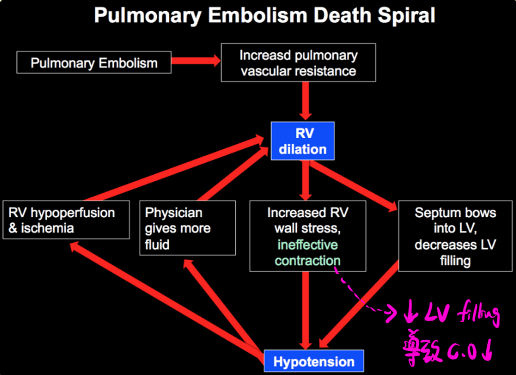
⭐️Pulmonary embolism➡︎increased PVR➡︎leads to RV dilation
- RV dilation pushes the septum toward the LV, reducing LV filling➡︎leading to lower cardiac output➡︎hypotension
- RV dilation increases RV wall stress, which in turn produces ineffective contraction➡︎hypotension
- Hypotension: leads to the following two situations
① RV hypoperfusion, then RV ischemia➡︎the RV dilates even more
② Because of the hypotension, the doctor gives more fluids➡︎the RV dilates even more
So the key to treatment➡︎in right-sided heart failure: forget ABC
➡️ A (Airway): does she need intubation?
- Avoid or delay intubation as much as possible
- PE patients usually die of circulatory failure, not respiratory failure➡︎intubation doesn't fix the underlying problem of PE, and is more likely to make the hemodynamics unstable
- If you really have to intubate➡︎turn the norepinephrine up and build a hemodynamic safety margin
- If she's an rTPA candidate, get the drug in fast first (delay intubation for now); after thrombolysis there's a chance oxygenation will gradually ↑
➡️ B (Breathing): does she need positive pressure ventilation?
- When a PE patient has poor oxygenation or excessive work of breathing→consider HFNC→maintain O2>95% and reduce work of breathing
➡️ C (Circulation): does she need a lot of fluid?
- Giving volume doesn't help much, and is even potentially harmful
- Current evidence in patients with acute PE plus RV dilation➡︎giving fluids doesn't help much
- The chance of a PE patient also being hypovolemic is low➡︎but these patients do respond to fluid➡︎approach: give 500 c.c NS→monitor the P’t; if hemodynamics stabilize or it doesn't work, stop
- Start norepinephrine early, before you've reached a stable blood pressure (please lower your threshold for using vasopressors)
- In the PE death spiral, when there's hypotension➡︎RV hypoperfusion and ischemia➡︎so don't put too many restrictions on starting vasopressors (low threshold)→↑B.P→↑RV perfusion
- If norepinephrine still can't maintain the B.P➡︎consider inhaled NO
Tips: Remember, in severe PE be bold and start vasopressors first to get the blood pressure up, rather than pouring in fluid first
If you're considering rTPA, remember to go through the rTPA check list (Fig.12) first for any contraindications

Case course: continuing with this case
When she was diagnosed, on NRM 15 L/min oxygen, her SaO2 was about 96%, her blood pressure was on the low side, and she was in shock.
We gave about 250 c.c of fluid, the blood pressure was still low, and I had the nurse go straight to a norepinephrine pump to get the pressure up first, no more fluid. Because keeping on pouring in fluid makes the RV dilate even more, which makes the RV ischemia worse and the shock more pronounced. Only by getting the pressure up does the RV hypoperfusion improve, and only then does the RV ischemia improve.
We gave the rTPA within about an hour of her arrival.
Because there was no ICU bed, after the rTPA she stayed in my hands for a full 12 hours (so painful XD), but that also meant I got to watch her clearly improve.
After we started the rTPA, her breathing pattern gradually improved, the oxygen came down from NRM 15 L/min to, in the end, N/C 3L/min, and the norepinephrine pump went from a sky-high dose to, in the end, off pump.
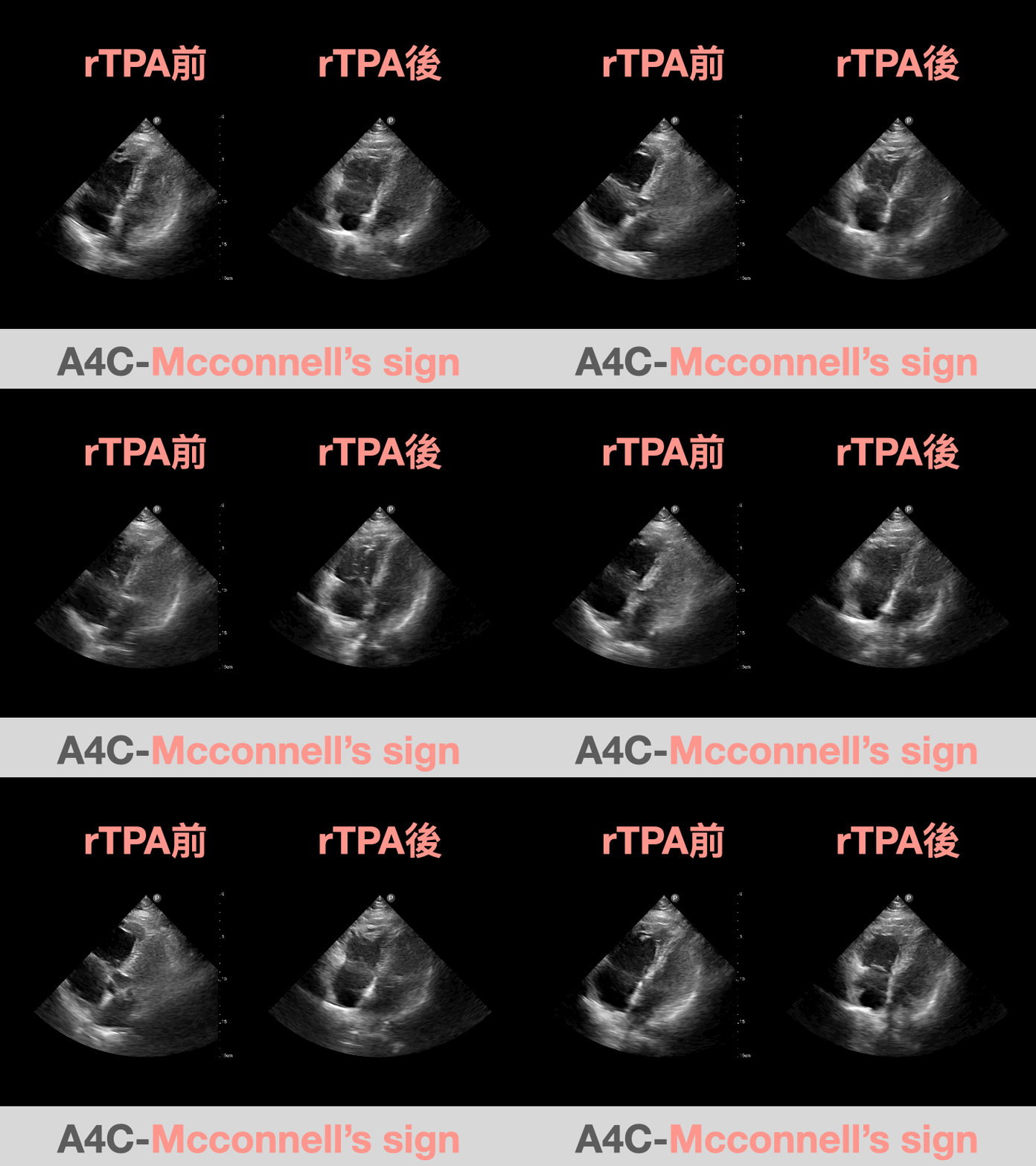
Fig.13 is the Heart POCUS I did again after the 2 hours of rTPA infusion had finished. The left side is before: an obvious McConnell’s sign. The right side is the A4C after rTPA: you can see the apical hyperkinesis has improved, and the RV free wall is moving much better too.
Key learning points:
- What are pathologic Q waves and septal q waves?
- Watch out: a big S wave in lead I may be caused by RV strain
- Which ECG features may indicate acute pulmonary embolism?
- What are the POCUS findings of acute PE?
- What are the key points in treating the crashing pulmonary embolism patient?
- Make good use of the simultaneity of the 12 lead ECG (vertical lines) and the consistency of a regular rhythm (horizontal lines); this often solves a lot of hard problems (remember to buy the steel ruler and Caliper)
Additional references: 1 2 4 3 5 7 8 9 10 6 11
Amal Mattu’s ECG Case of the Week — December 21, 2020 — ECG Weekly — link ↩︎ ↩︎
Amal Mattu’s ECG Case of the Week — April 11, 2022 — ECG Weekly — link ↩︎ ↩︎
Amal Mattu’s ECG Case of the Week — June 27, 2022 — ECG Weekly — link ↩︎ ↩︎
Digby, G. C., Kukla, P., Zhan, Z.-Q., Pastore, C. A., Piotrowicz, R., Schapachnik, E., Zareba, W., Bayés de Luna, A., Pruszczyk, P., & Baranchuk, A. M. (2015). The Value of Electrocardiographic Abnormalities in the Prognosis of Pulmonary Embolism: A Consensus Paper. Annals of Noninvasive Electrocardiology, 20(3), 207–223. https://doi.org/10.1111/anec.12278 ↩︎ ↩︎
Dr. Smith’s ECG Blog: A woman in her 50s with shortness of breath — link ↩︎ ↩︎
Co, I., Eilbert, W., & Chiganos, T. (2017). New Electrocardiographic Changes in Patients Diagnosed with Pulmonary Embolism. __The Journal of Emergency Medicine__, __52__(3), 280–285. https://doi.org/10.1016/j.jemermed.2016.09.009 ↩︎ ↩︎
ECG Case 123 • LITFL • ECG Top 100 Self-Assessment Quiz — link ↩︎ ↩︎
Kosuge, M., Kimura, K., Ishikawa, T., Ebina, T., Hibi, K., Kusama, I., Nakachi, T., Endo, M., Komura, N., & Umemura, S. (2007). Electrocardiographic differentiation between acute pulmonary embolism and acute coronary syndromes on the basis of negative T waves. The American Journal of Cardiology, 99(6), 817–821. https://doi.org/10.1016/j.amjcard.2006.10.043 ↩︎ ↩︎
Raunio, H., Rissanen, V., & Lampainen, E. (1976). Significance of a prominent S wave in leads I and V6 in the electrocardiograms of middle-aged and elderly hospital patients. Annals of Clinical Research, 8(6), 347–358. ↩︎ ↩︎
Eight pearls for the crashing patient with massive PE — link ↩︎ ↩︎



Thoughts on this case? Leave a comment, a like, or a reaction below.
comments powered by Disqus