Translated from the original Traditional Chinese post. Read the Chinese original →

IHCA → Pulmonary embolism, or something else?
Let's look at an interesting case.
85-year-old woman with a history of cancer. On arrival: dyspneic, hypotensive, confused.
ECG interpretation (see the image above): unstable baseline
Rate: 120
Rhythm: Sinus
Axis: RAD
New RBBB (not on her prior ECG), 1st AVB
The baseline is unstable, but there seems to be STD over V2~V6 and I/aVL (maximal in V3~V4). It would be clearer if we could repeat it, but given how sick she was, this may already be as clean as it gets. STE over aVR and III.
In V1 it's hard to tell whether it's STE or STD. If it's STE:
then right precordial lead STE (meaning V1, V2, aVR, ±III) + RAD should make you specifically consider three DDx: hyper-K, PE, and Na channel blocker toxicity
⭐️The case continues
After that, because her breathing was poor and her blood pressure was low, she was close to collapse.
During the workup, PE was still the top diagnosis to rule out. (Clinical picture; ECG: RAD, new RBBB; POCUS: what looked like RV dilation but no D-sign)
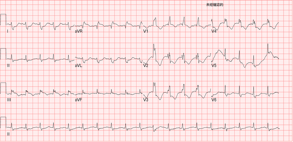
This is the ECG 50 minutes later (Fig.1), with almost no baseline wander.
RAD, RBBB, STE over III and aVR, STD over V2~V6 (maximal at V3~V4) and I/aVL. Here we can be quite sure the maximal precordial lead is V3~V4, and the STD is excessively discordant.
In an RBBB pattern, it's common to see TWI and mild STD in V1~V3 (if any at all), but the STD does not exceed 1 mm. So going beyond 1 mm means some force is pulling the ST segment down1.
➡️What force? A posterior wall OMI (occlusion of the vessel supplying the posterior wall).
Also, the DDx for aVR STE + diffuse STD is very long.
Some of it is vascular, some of it isn't.
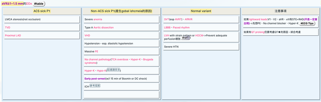
Bottom line: seeing aVR STE is like looking into a wastebasket. Lots of diagnoses live in there. If the patient has ACS symptoms plus aVR STE, then yes, you do need to consider LM, TVD, or proximal LAD disease.
But far more often there are no ACS symptoms and all you've got is aVR STE. In that situation, do not spinal-reflex your way into calling it one of the vascular problems above. Think carefully about the possible causes.
One more thing to note: the ESC 2020 NSTE-ACS guideline says that very high-risk non-STEMI patients are recommended to get emergent PCI (Fig.2, lower-left table) within 2 hours
In the table you can see that, besides the ones we all know (hemodynamic instability, VT/VF, ischemia-related HF, refractory ischemic pain, cardiogenic shock), having STD > 1 mm in at least 6 leads + aVR STE also counts as a very high-risk patient.
Of course, this table only applies if the patient actually is a non-STEMI patient! As for whether this one is, read on~~~~

The CV man came down and put her on PCPS to support her vital signs so we could continue the workup. While on PCPS, she completely collapsed and CPR was started.
Question: if at this point we strongly suspect the collapse or peri-arrest is from a pulmonary embolism, how do we manage it?
- For PE with collapse, aggressive CPR + immediate rTPA (50 mg push) → one study showed 35% survival to discharge (MAPPET study)2
⭐️The case continues again
Once she was on the mini Dr. Yeh (PCPS; ECMO's Mandarin name, Ye-ke-mo, sounds like a Dr. Yeh), she got ROSC and gradually stabilized. She then went for a chest CTA, which showed no pulmonary vascular occlusion at all.
Later, her TnI from the ED came back at about 30 ng/ml.
She went up to the ICU first. Fig.3 is the ECG after ICU admission (about 4.5 hrs from the ER).
Same STD over V1~V6 (maximal at V3~V4) and RBBB, but the rate has slowed down.

Here's one small but important point. Look back at the aVR STE DDx table above: did you notice SVT and AfRVR listed among the normal variants?
Clinically, when we see PSVT or AfRVR running very fast, oxygen demand/supply mismatch makes a tachycardia-related global ischemic ECG pattern likely.
Question: if you see a very fast heart rate together with multiple-lead STD + aVR STE, how do you tell whether it's a posterior wall OMI or diffuse subendocardial ischemia?
Podcast 128 - Who Gets A Right-Sided ECG? w/ Dr. Stephen Smith & Tom Bouthillet
In this podcast, the host Tom Bouthillet asks Dr. Smith (start listening at 16:41):
- With a tachycardia like AfRVR, the rate is so fast that an ECG pattern of aVR STE with diffuse STD shows up. Should you bring the H.R down first before deciding whether it's a posterior OMI or subendocardial ischemia? ➡ Stephen Smith says bring the H.R down first (to remove the tachycardia-related ischemic ECG pattern as a factor)
- If the H.R comes down and the subendocardial ischemia goes away, but you still see maximal STD in V1~V4, that looks more like a posterior OMI.
- Also, the role of bedside echo ➡ in subendocardial ischemia it will be completely normal (because the epicardial vessels are normal); in a posterior OMI you may see a basal inferolateral wall RWMA (Segment 5 of the 17 segments). If the echo looks like it's moving well, then once the H.R comes down, aVR STE + multiple-lead STD caused by subendocardial ischemia should improve.
Want to see more cases like this to sharpen your skills?3
A man in his early sixties with palpitations
Actually, the paper says it's Segment 4 that is the so-called posterior wall (Fig.4)4

Tips: if a fast H.R is causing multiple-lead STD + aVR → bring the H.R down first (if you still see maximal STD over V1~V4 after the H.R comes down → think posterior OMI)
Tips: echo can also help distinguish posterior OMI from subendocardial ischemia
⭐️The case continues
Look at Fig.3: the heart rate has already slowed, and there is maximal STD in V1~V4. So either way, going by Dr. Smith's latest research paper (coming out soon in the Journal of the AHA):

Either way, you must think posterior OMI first, until proven otherwise. The point here is that whenever you see maximal STD over V1~V4, no matter how small the amplitude, just guess posterior OMI first and you'll be right (highly specific).
Her TnI 4 hours later shot up to 250 ng/ml (large myocardial infarction). She then went to the cath lab for PCI 5 hours later. The CAG is shown below in Fig.5, showing LCx critical stenosis and an RCA CTO
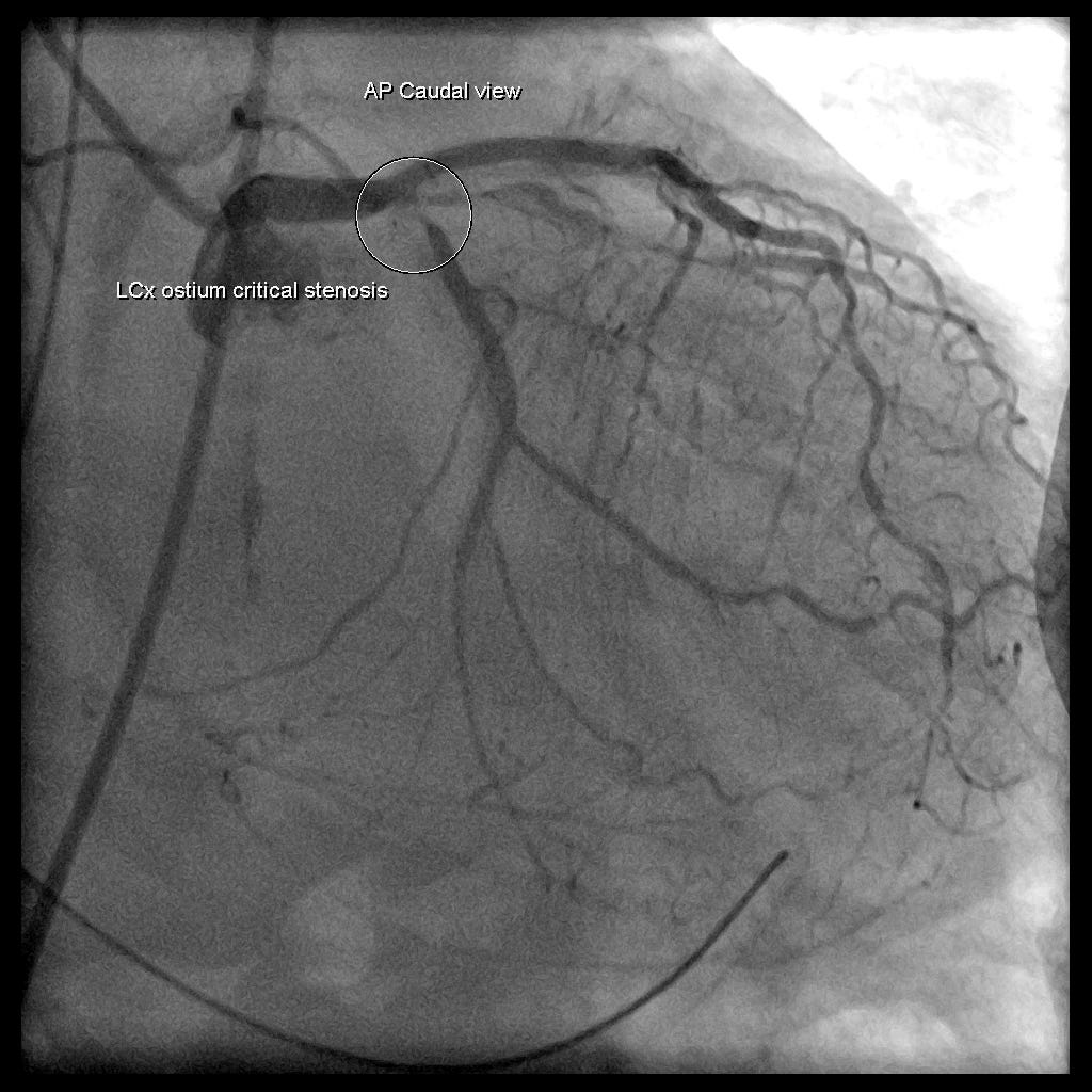
So in this case the collapse was caused by a large myocardial infarction from LCx critical stenosis, not by a pulmonary embolism.
Let's look at the post-PCI ECG (Fig.6), and you'll see that the LCx really was the culprit lesion; we're not just saying that.
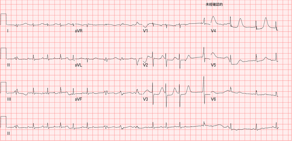
The one above is the post-PCI ECG. What do we see → the T waves got bigger and upright in the precordial leads.
First we need to know that a reperfusion rhythm shows up in two situations: see the reperfusion ECG changes at the bottom of Fig.7
- The vessel opened on its own
- Someone else opened it (with PCI or thrombolytic agents)
A normal reperfusion rhythm first shows biphasic TWI, which then becomes deep TWI → remember Wellens' syndrome? Wellens' syndrome is exactly the ECG pattern of a reperfusion rhythm.
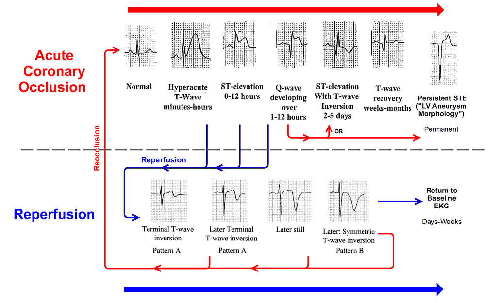
So what does a reperfusion rhythm look like in a posterior OMI?
V1~V4 will show bigger, upright T waves (which, mirrored onto the posterior wall, is that deep-TWI reperfusion rhythm). So, do you see the huge T waves in V2~V4 → this is also called posterior Wellens' syndrome.
Be careful here not to confuse it with De Winter's T waves. You canrefer to this post, an article I wrote on telling them apart5.
⭐️Key points of this post:
- Checking for ischemia in an RBBB morphology → the trick: there should be no ST deviation at all (except possibly mild STD and TWI in V1~V3), and the STD should not exceed 1 mm
- How to diagnose RBBB + posterior OMI → excessively discordant STD in V1~V4 (that is, STD of more than 1 mm) should make you consider it
- If there's also tachycardia-related aVR STE + multiple-lead STD → bring the H.R down first to remove the subendocardial ischemia caused by the fast rate; if, after that, you still see maximal STD in V1~V4 → remember to consider posterior OMI
- If you see multiple-lead STD in the precordial leads and can't tell subendocardial ischemia from posterior OMI → bedside echo and posterior leads can help tell them apart. But posterior leads can be false-negative (that is, there really is a posterior OMI, but because of low voltage the STE in the posterior leads isn't obvious, so it's misjudged as absent).
- In the end, I still think that with this case it was really hard for me to tell, in the first moment, whether this was PE or AMI. Fortunately, the CV man strongly backed PCPS, which is what made the later workup and diagnosis possible. That said, this case's ECGs taught me a lot.
Additional references: 2 1 4 3 5
Dr. Smith’s ECG Blog: Posterior ST Elevation MI in the Setting of Right Bundle Branch Block, with Posterior Leads V7-V9 — link ↩︎ ↩︎
Kasper, W., Konstantinides, S., Geibel, A., Olschewski, M., Heinrich, F., Grosser, K. D., Rauber, K., Iversen, S., Redecker, M., & Kienast, J. (1997). Management Strategies and Determinants of Outcome in Acute Major Pulmonary Embolism: Results of a Multicenter Registry. __Journal of the American College of Cardiology__, __30__(5), 1165–1171. https://doi.org/10.1016/S0735-1097(97)00319-7 ↩︎ ↩︎
Dr. Smith’s ECG Blog: A man in his early sixties with palpitations — link ↩︎ ↩︎
Bayés de Luna, A., Wagner, G., Birnbaum, Y., Nikus, K., Fiol, M., Gorgels, A., Cinca, J., Clemmensen, P. M., Pahlm, O., Sclarovsky, S., Stern, S., Wellens, H., & Zareba, W. (2006). A New Terminology for Left Ventricular Walls and Location of Myocardial Infarcts That Present Q Wave Based on the Standard of Cardiac Magnetic Resonance Imaging: A Statement for Healthcare Professionals From a Committee Appointed by the International Society for Holter and Noninvasive Electrocardiography. __Circulation__, __114__(16), 1755–1760. https://doi.org/10/d4gnjk ↩︎ ↩︎
[Is This Posterior Wellens' Syndrome or De Winter?] (in Chinese) — Tsao Jian-hsiung — Medium — link ↩︎ ↩︎



Thoughts on this case? Leave a comment, a like, or a reaction below.
comments powered by Disqus