Translated from the original Traditional Chinese post. Read the Chinese original →
Chest pain: is it Wellen?
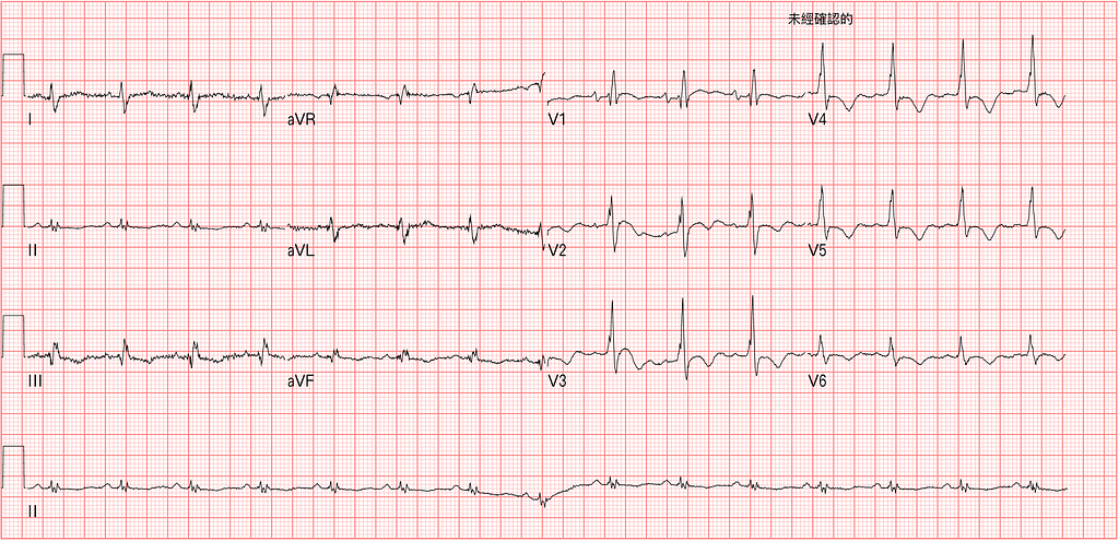
A 70-year-old man with chest pain for three days. At night it hurt so much he couldn't sleep. Past history of HCC treated with transarterial embolization, still under treatment.
On closer questioning, his upper abdomen was also uncomfortable, and he was a tiny bit short of breath, but chest pain was the main thing.
Going further: rate about 90, P waves upright in Lead I/II/aVF and inverted in aVR, so it's sinus rhythm.
Axis change. In Lead I the S wave > R wave → so RAD
PR interval normal, rSR' pattern in V1~V3, QRS < 3 small boxes, so ICRBBB.
The more obvious abnormality is TWI in the precordial leads (V1~V6), plus TWI in the inf. leads too. Q wave in III.
ECG read done. Let's look at a few questions~~~
1️⃣ ➡︎Is the TWI in the precordial leads Wellen's wave?
Why ask this? Because it's the question our knee-jerk reflex jumps straight to?
2️⃣➡︎If it's not Wellen's wave, what else could give this kind of TWI?
Is the TWI in V1~V6 Wellen's wave? We have to know what Wellen's wave is before we can make that diagnosis.
When we usually say a patient may have Wellens' syndrome, we mean seeing biphasic TWI or deep TWI in V2~V3, seen after ACS symptoms resolve.
That is, the patient first has ACS symptoms, then the symptoms resolve, and the ECG done during that pain-free period shows these two kinds of TWI in V2~V3. Then we can call it Wellens' syndrome.
So what does Wellens' syndrome mean?
Clinically, it means a high risk of an extensive ant.wall MI within the next few days to few weeks.
So why do these two kinds of TWI appear?

First, let's look at Fig.1. The top of this figure covers the ECG changes of ACO (Acute coronary occlusion). The bottom covers the typical ECG changes when reperfusion happens before infarction. We'll get to that later.
So what causes reperfusion? Spontaneous reperfusion (it opens on its own), giving rTPA, or opening it with PCI.
This paper describes that about 20% of STEMI patients have spontaneous reperfusion before PCI1. There's also a really important concept here: coronary occlusion is dynamic. In the IRA (infarct related artery), endogenous tPA and plasmin can dissolve the thrombi, leading to spontaneous reperfusion.
This post on Smith's ECG Blog says that 19% of STEMIs have spontaneous reperfusion with TIMI flow-32.
Tips: Vessel occlusion is dynamic, so spontaneous reperfusion can happen

Now let's talk about the typical ECG changes after reperfusion, which include Pattern A and Pattern B in Fig.2.
Pattern A is called terminal TWI, i.e., biphasic TWI, and Pattern B is deep TWI. Both A and B are reperfusion T waves.
Usually Pattern A evolves into Pattern B. And the more obvious this reperfusion evolution, the more it indicates that a certain amount of myocardium was salvaged3. In other words, the depth of the TWI correlates with viable myocardium4.
Wellens' syndrome means the vessel was occluded and then suddenly opened (spontaneous reperfusion) before infarction. So reperfusion T waves appear. Because it's a syndrome named after Wellen, at the time it specifically meant TWI in V2~V3. Of course reperfusion T waves don't have to be in V2~V3; they can show up in the lateral leads, high lateral leads, or inf. leads too. It just depends on whether the artery supplying those territories has reperfused.
Let's look at the diagnostic definition, as described in the paper by Rhinehardt et al5
- Deeply inverted or biphasic T waves in V2–3 (may extend to V1–6)
- ECG pattern present in pain-free state → TWI appears when symptoms have resolved
- Isoelectric or minimally-elevated ST segment (< 1mm)
- No precordial Q waves →precordial Q waves mean infarction has already happened, which doesn't fit the definition of ECG changes before obvious infarction
- Preserved precordial R wave progression →PRWP may mean infarction has already happened
- Recent history of angina → a prior episode of typical chest pain lasting at least 20 minutes
- Normal or slightly elevated serum cardiac markers
There are a few key points in this definition for diagnosing Wellens' syndrome →the ECG must be done while symptoms have resolved, and there must be no Q waves in the precordial leads and preserved R wave progression. These have to be met before you can call it Wellen's syndrome.
So seeing TWI in the precordial leads does not automatically make it Wellens' syndrome.
This ECG was done while the patient was symptomatic. Even though there's precordial TWI, it can't be called Wellens' syndrome.
So what else causes TWI in the precordial leads?
Smith's ECG Blog says that most TWI is non-specific6.
Also, RV strain can cause TWI in the R't precordial leads ± inf. leads.
Besides the ECG findings above, RV strain can also show the following changes:
- Tall R wave in V1 (R/S >1) →this exists in only 1% as a normal variant, so it's not common in the ER. If you see it, dig into the cause
- RAD
- ST changes (especially STE in the rightward leads) → rightward leads means V1/V2/aVR/III
Let me say a bit more about the DDx of the first point, tall RV1 (Fig.3)
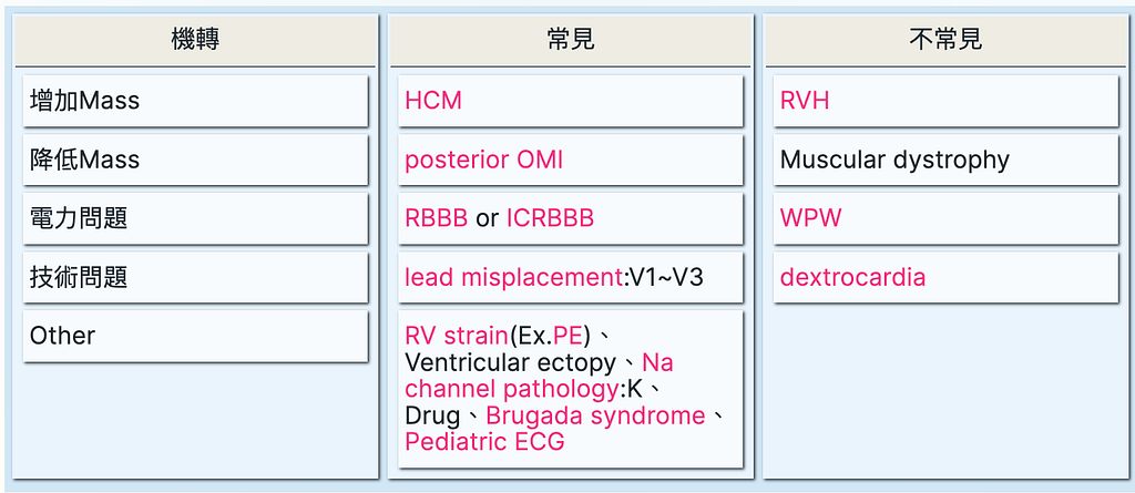
I split it into four parts to remember. Remembering the more common ones is enough to fight the monsters.
Only the "other" group is hard to remember: RV strain, ventricular ectopy, Na channel pathology (hyper-K, Na channel blocker toxicity, and Brugada syndrome), and pediatric ECGs, plus of course normal variant.
Here's another mnemonic: R-WAVED

Another key point here →the ECG can identify RV strain, but it can't tell you the cause of the RV strain➡ possible causes are PE, asthma/COPD, hypoxic vasoconstriction from pneumonia, pulmonary HTN.
RV strain means the RV is being stretched taut and distended. So when you see an RV strain ECG pattern, think about whether something is wrong with the pulmonary vasculature: an occlusion, or pulmonary hypertension, that in turn causes the RV strain.
So in this case, with RAD and TWI in the R't precordial leads/III/aVF, we definitely have to consider RV strain.
Looking at the ECG alone, when we see TWI in the precordial leads, can we use ECG morphology to tell whether it's ACS (Wellen) or PE?
There are key results from a few studies we can draw on.
The first is a paper by Kosuge et al published in 20077. It has an important conclusion: if the diagnosis comes down to ACS vs. PE, then an ECG with TWI in the precordial leads together with TWI in III/V1 should make you strongly consider PE.
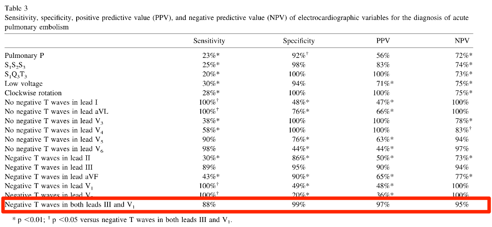
What Fig.4 is saying is that when it shows up in both III/V1, 88% of PE look like this, and only 1% of ACS do.
Another paper by the same Kosuge research team8, published in 2016, uses where the peak TWI sits in the precordial leads to tell them apart.
Its conclusion →Peak negative TWI in V1~V2, when used to diagnose PE ➡ 95% sensitivity, 89% specificity, 89% PPV, 95% NPV.
You can also look at the shape of the TWI to tell ACS from PE (Fig.5).

Look at Fig.5: the 5 cases in the top row are all PE, and the 5 cases in the bottom row are all ACS-Wellen.
Is there a difference? Yes!!!!!!
In PE the TWI is convex STE that then curves downward like a roller coaster. In ACS-Wellen the TWI drops steeply and is sharper.
Dr. Smith describes this kind of PE TWI on his blog and calls it Domed TWI (dome-shaped TWI)9.
When you look at it and just don't feel it, burn Fig.5 above into your brain. Pull it up and worship it every so often. You'll definitely get the feeling~~~~
So if we apply the three methods above for telling ACS vs. PE TWI apart to the case I saw, how many fit?
- TWI in the precordial leads together with TWI in III/V1 →(O)
- Peak TWI in V1~V2 →(X) (in this case the peak TWI was in V4~V5)
- TWI morphology →(O) (the TWI in this case was Domed TWI)
Here goes~~~ the moment of truth~~~
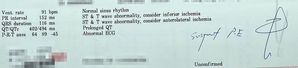
After seeing the ECG, I wrote down suspected PE right away. And ordered a Chest CTA.
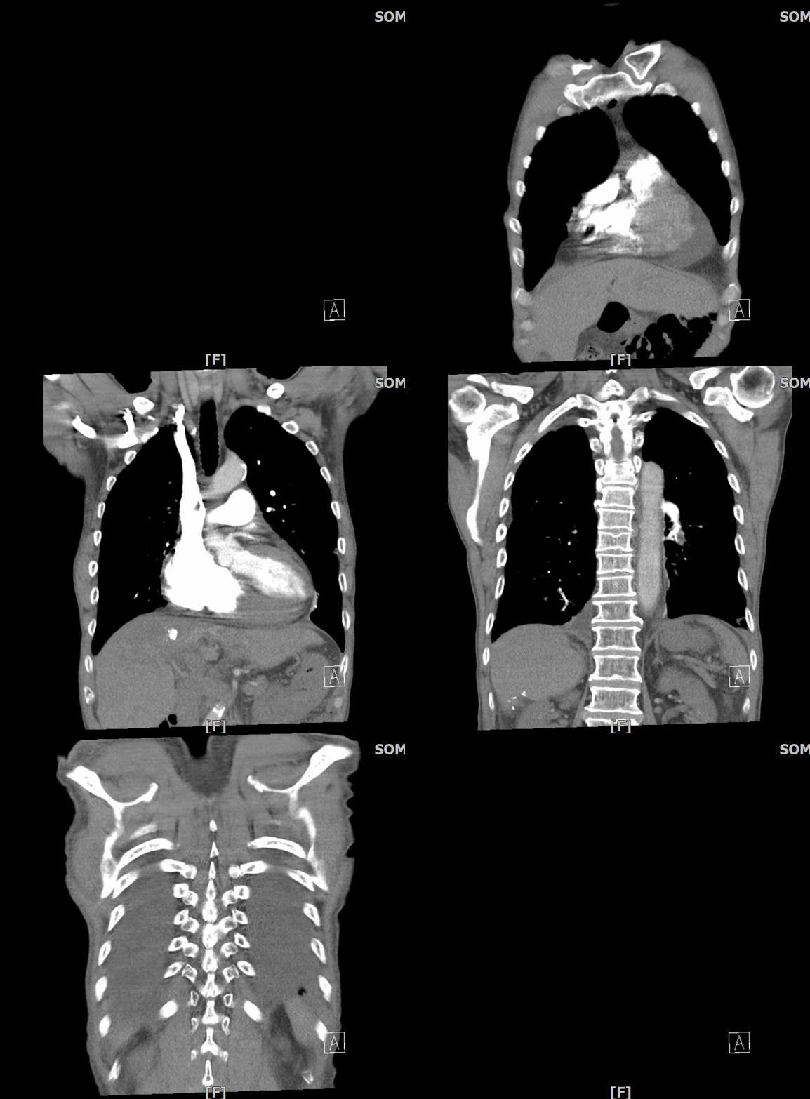
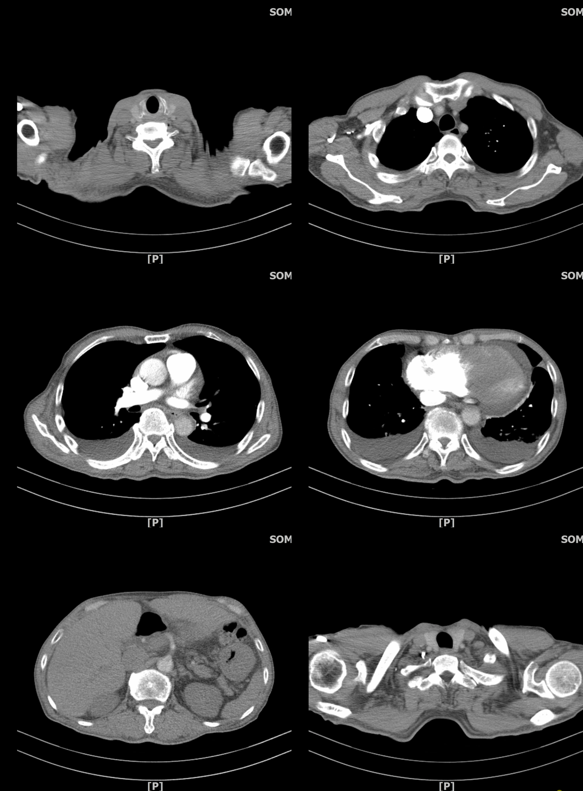
Chest CTA showed bilateral pulmonary vessel filling defects, and a filling defect in the RV too. Bilateral pulmonary infarction~~~~
At the time I also did a bedside echo on the patient.
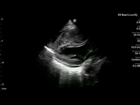
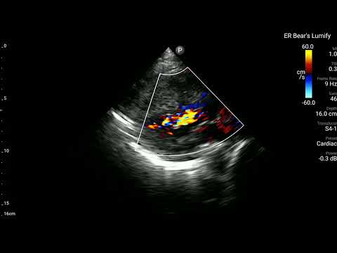
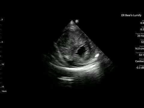
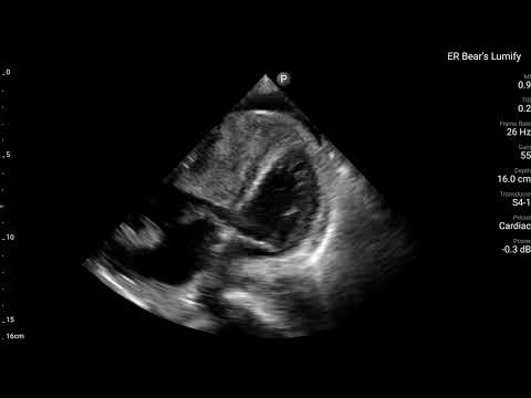
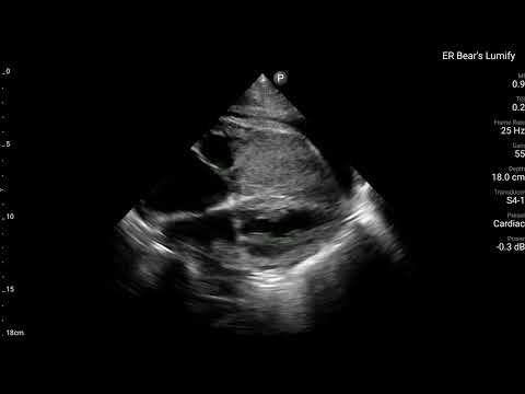
You can clearly see a whole mass lesion in the RV. Turning on color flow in PLAX, there's no obvious flow in the RV either. In the PSAX view you can clearly see RV dilation with a D-sign. The A4C view shows even more clearly that the RV is packed with something. Whether it's tumor or thrombus, it has basically already caused RV strain (obvious RV dilation). Which in turn produced the changes on the ECG.
Take-home points:
- What is Wellens' syndrome? What does it mean?
- What are the ECG findings of RV strain?
- What conditions can cause a tall RV1?
- The ECG can identify RV strain, but it can't tell you the cause of the RV strain
- Can we use ECG morphology to tell whether it's ACS (Wellen) or PE?
Additional references: 1 2 3 4 5 6 7 8 9
Fefer, P., Hod, H., Hammerman, H., Boyko, V., Behar, S., Matetzky, S., & Acute Coronary Syndrome Israeli Survey (ACSIS) 2006 Study Group. (2009). Relation of clinically defined spontaneous reperfusion to outcome in ST-elevation myocardial infarction. __The American Journal of Cardiology__, __103__(2), 149–153. https://doi.org/10.1016/j.amjcard.2008.08.050 ↩︎ ↩︎
Dr. Smith’s ECG Blog: ST Depression Maximal in V1-V4 and Angio shows 3 Vessel Disease. Is it posterior? Which is the culprit? — link ↩︎ ↩︎
Dr. Smith’s ECG Blog: A man in his 30s with greater than 12 hours of chest pain — link ↩︎ ↩︎
Dr. Smith’s ECG Blog: Is the LAD really completely occluded when there are de Winter’s waves? — link ↩︎ ↩︎
Rhinehardt, J., Brady, W. J., Perron, A. D., & Mattu, A. (2002). Electrocardiographic manifestations of Wellens’ syndrome. __The American Journal of Emergency Medicine__, __20__(7), 638–643. https://doi.org/10.1053/ajem.2002.34800 ↩︎ ↩︎
Dr. Smith’s ECG Blog: Why we need continuous 12-lead ST segment monitoring in Wellens’ syndrome — link ↩︎ ↩︎
Kosuge, M., Kimura, K., Ishikawa, T., Ebina, T., Hibi, K., Kusama, I., Nakachi, T., Endo, M., Komura, N., & Umemura, S. (2007). Electrocardiographic differentiation between acute pulmonary embolism and acute coronary syndromes on the basis of negative T waves. __The American Journal of Cardiology__, __99__(6), 817–821. https://doi.org/10.1016/j.amjcard.2006.10.043 ↩︎ ↩︎
Kosuge, M., Ebina, T., Hibi, K., Tsukahara, K., Iwahashi, N., Maejima, N., Akiyama, E., Umemura, S., & Kimura, K. (2016). Simple electrocardiographic criteria for discriminating between acute pulmonary embolism and acute coronary syndrome. __Journal of the American College of Cardiology__, __67__(13_Supplement), 528–528. https://doi.org/10.1016/S0735-1097(16)30529-0 ↩︎ ↩︎
Dr. Smith’s ECG Blog: Are these Wellens’ waves? — link ↩︎ ↩︎



Thoughts on this case? Leave a comment, a like, or a reaction below.
comments powered by Disqus