Translated from the original Traditional Chinese post. Read the Chinese original →
Besides STE, Which Other ECG Signs Can Mean an Occluded Artery? (Introduction)
Let's look at a few ECGs first, then come back to the question above~~
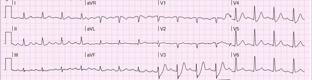

First, let's ask ourselves: do these ECGs meet STEMI criteria?
If not, would we Call CV man right away?
And if it's the middle of the night and you Call CV for an ECG pattern like this, are you going to get yelled at?
We also have to ask ourselves: if an ECG doesn't meet STEMI criteria, how do you tell there's an occluded artery? What literature is out there right now that tells us an ECG pattern like this means occlusion myocardial infarction (Occlusion MI →OMI)?
Whoa~~~~ that's way too many questions XD
Let me sum up the questions above. They boil down to a few key points.
First: is diagnosing myocardial infarction with STEMI criteria reliable?
Second: besides STEMI criteria, which other ECG patterns can help diagnose myocardial infarction?
Are STEMI criteria reliable? For diagnosing myocardial infarction, yes (specificity is high enough)…… but the sensitivity is terrible.
This paper1 shows that applying STEMI criteria to the first ED ECG gives a sensitivity of only 35%. What does that mean? It means 65% of myocardial infarctions can't be diagnosed by STEMI criteria.
Is it really only 35%? Another paper2 got to 50% sensitivity, but that's still pretty underwhelming~~~
So if we insist that an MI patient must meet STEMI criteria before we send them for cath, the problem is that we'll miss a lot of myocardial infarctions up front. The patient may still end up getting cath eventually, but Time is muscle. The longer it drags on, the more of the patient's myocardial cells die, and the longer the heart failure and cardiac rehab that follow.
Besides STEMI criteria, which other ECG patterns can help diagnose myocardial infarction?
Before answering that, let's look at what some of the literature says.
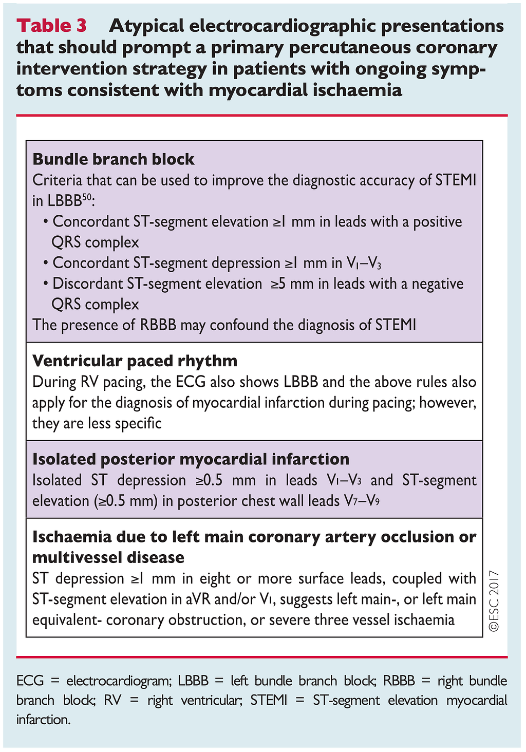


Let's start with the left side of Table 1, the table from the ESC 2017 STEMI guideline. It describes the listed situations as atypical ECG presentations of myocardial infarction for which the cath lab should be activated
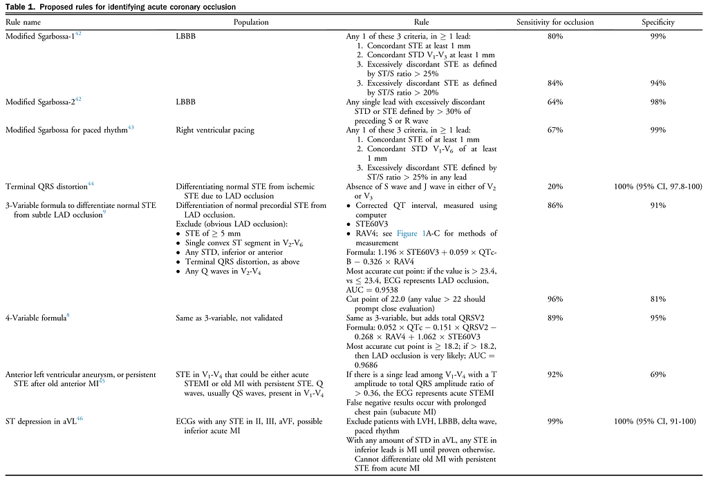
The middle of Table 1 comes from this paper3, which describes the listed situations as ones where ACO (Acute coronary occlusion) needs to be considered
The right side of Table 1 comes from this paper4, which describes what counts as an OMI ECG finding
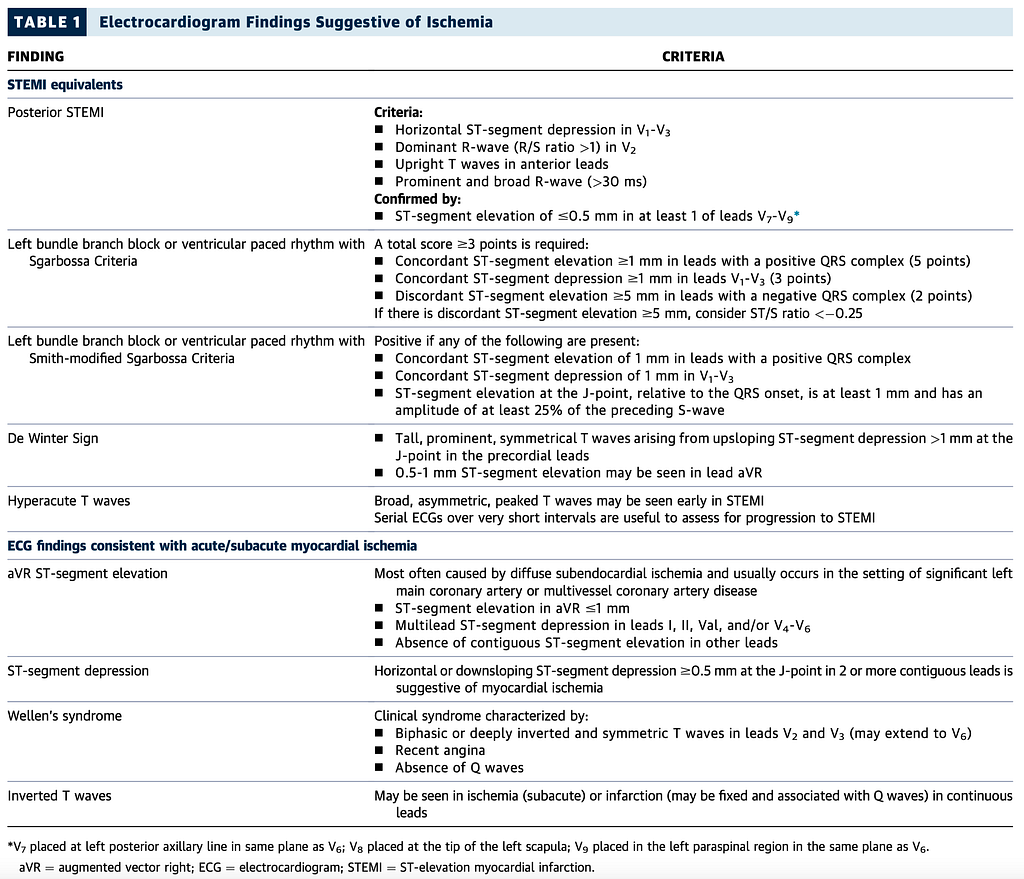
The left side of Table 1 Part II is a table from the ACC expert consensus on acute chest pain in the ED, published in October 20225. It covers STEMI equivalents and which ECG patterns may come with acute/subacute myocardial ischemia (the original table has a few small mistakes, see if you can spot them XD)
The right side of Table 1 Part II is from Dr. Smith's latest paper6, which lists which ECG patterns carry a high risk of ATO (acute total coronary occlusion).
That's a hell of a lot…….
Yep, that's the reality. Since STEMI criteria are so insensitive for diagnosing MI, more and more ECG patterns are only going to get published saying this situation could be MI too. Just like our Tintinalli pillow, which keeps getting taller the more we sleep on it XD
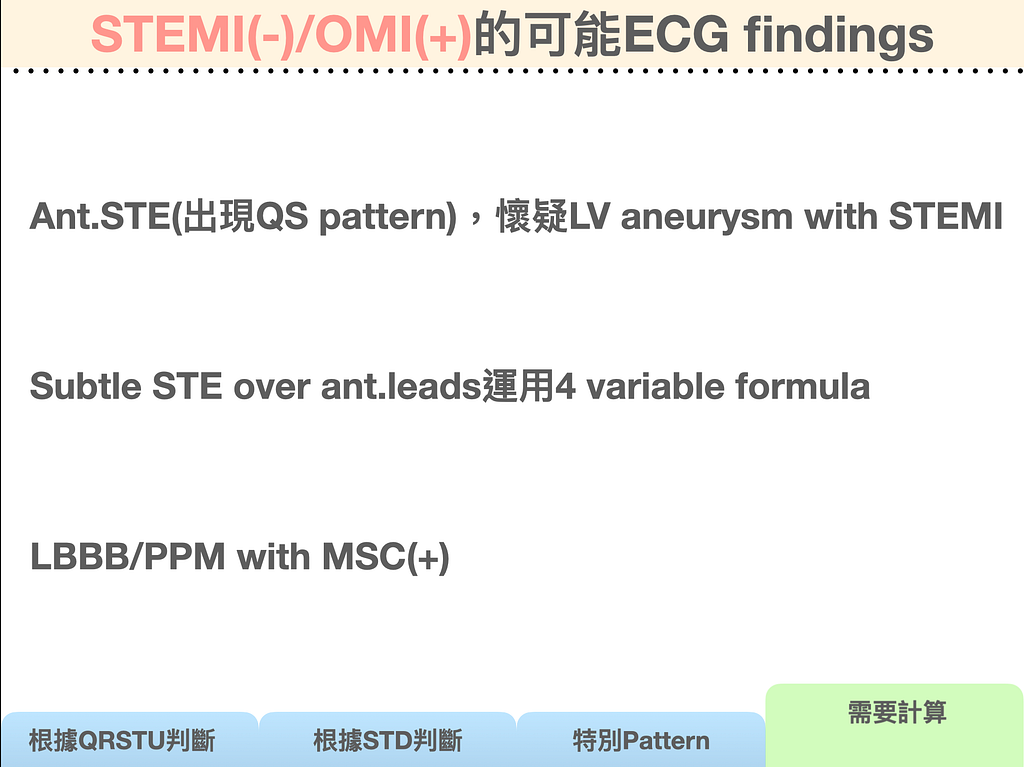
I've taken the ECG patterns that Amal mattu and Dr. Smith have written about in recent years as possibly coming with MI, sorted them into the following major categories, and tidied them up a bit so they're easier to follow. I gave a talk on this at Dalin Tzu Chi Hospital on October 31 (250 slides, I nearly coughed up blood). If you're interested, you can……..


In case some readers still don't know what Occlusion MI (OMI) is, here's a quick explanation.

Simply put, STEMI/NSTEMI is distinguished by the ECG, while OMI/NOMI is diagnosed by the pathology of the artery. In other words, an MI with an occluded artery is called OMI. Of course, assessing whether the artery may be occluded ultimately comes down to the cath result. But before cath, the ECG really is one of the most important tools for diagnosing MI, and echo and cardiac enzymes (Troponin-I, Troponin-T) can also help.
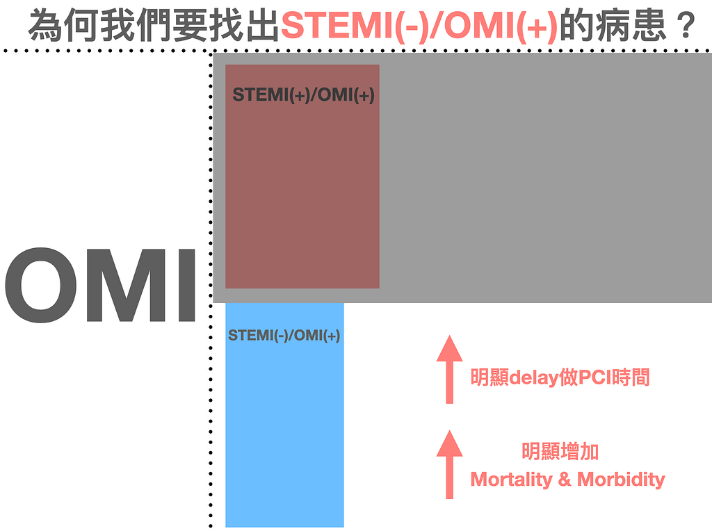
So the two most important boxes in Fig.5 are STEMI(+)/OMI(+) and STEMI(-)/OMI(+). Both are type 1 MI, both caused by plaque rupture leading to thrombosis and an occluded artery. They're also the two groups most worth taking to cath to save myocardial cells.
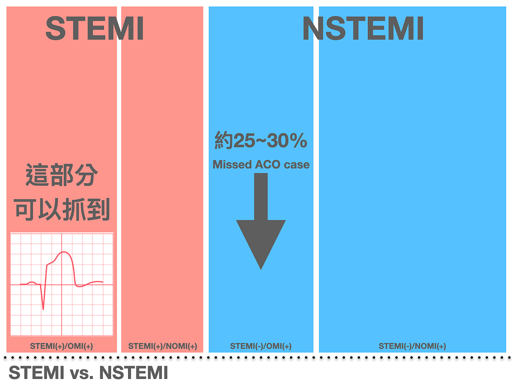
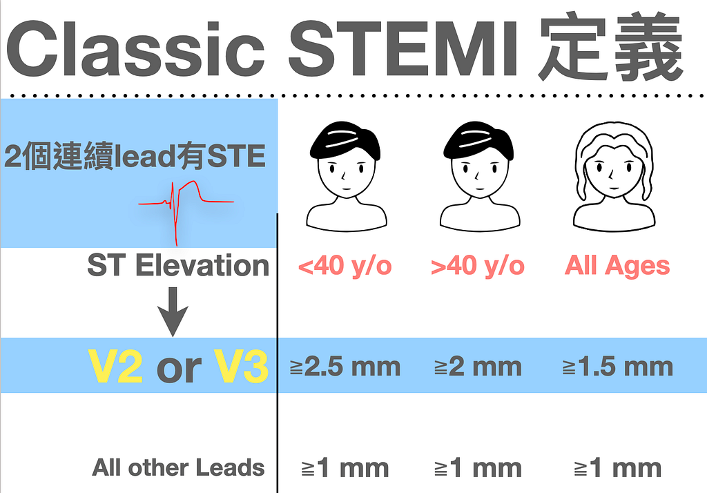
Let's look at Fig.6. It shows that STEMI(+)/OMI(+) can be caught with STEMI criteria. Don't know the basic definition of STEMI criteria? (See Fig.7)
Whatever you do, remember that of the 12 leads, V2-V3 are different from the other 10 leads: for V2-V3 you have to look at age and sex.
Now on to the STEMI(-)/OMI(+) box in Fig.6. Among NSTEMI patients, statistically 25–30% are missed ACO patients.
Let me explain what that means. In the past, if an ACS patient was diagnosed with NSTEMI (doesn't meet STEMI criteria, but biomarkers are up and there's evidence of myocardial ischemia +), we'd usually consult the CV man, and there'd usually be a few possible answers:
- Admit to the ICU for observation
- Admit to the ward for observation
- Cath right away
Usually the patient gets admitted for observation, and a few days later a cath is scheduled, but in 25–30% of patients, when they go in with the cath, the artery is occluded. These are the so-called STEMI(-)/OMI(+) patients: the artery is solidly, completely plugged up, yet the cath to open it happens days late.
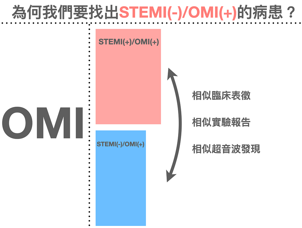
Someone might ask: the patient got the cath in the end anyway!!! So why do we need to find STEMI(-)/OMI(+) patients? What difference does it make?
It makes a difference~~~ it really does.
This paper7 compared the STEMI(+)/OMI(+) and STEMI(-)/OMI(+) groups. Both had similar clinical presentation/lab results and echo findings. But in the STEMI(-)/OMI(+) group, cath was clearly delayed, and compared with the STEMI(+)/OMI(+) group, mortality and morbidity were clearly higher (see Fig.8).
Another paper8 reports NSTEMI with ATO (acute total coronary occlusion) at about 34%. All-cause mortality, recurrent MI, and cardiogenic shock were all clearly higher in the NSTEMI with ATO group than in the NSTEMI without ATO group.
Picture an NSTEMI with ATO patient: the whole artery is occluded, but…… we wait days before opening it. Just thinking about it, who knows how much myocardium has died. And the studies do show that the NSTEMI with ATO group, compared with the group without ATO, has clearly worse CV outcomes.
So back when the OMI concept hadn't caught on yet (has it caught on now?), the ESC 2020 NSTEMI guideline also said patients with very high risk features need PCI within < 2 hr (see Fig.9).

The lower left corner of Fig.9 lists many situations in which cath is recommended within 2 hours. Apart from aVR STE + multiple leads STD, it says almost nothing about what the ECG pattern has to look like before the patient can go to cath. That means the ESC guideline also knows STEMI criteria aren't the only criterion for diagnosing MI: there are plenty of cases where the artery clearly is occluded, or is progressively occluding, but you just don't see anything that meets STEMI criteria. That's why it recommends cath for patients with a Very high risk feature. Note that it's Very high risk feature, not "Very high risk ECG feature".
The 2020 ESC NSTEMI guideline recommends PCI within <2 hours. When I teach, I often ask medical students to guess: in real-world clinical practice, what proportion actually get PCI within <2 hours?
Some say 60%
Some say 80%
All wrong~~~~ in actual clinical practice, it's not high.
Let's see what Dr. Smith wrote in a Twitter post this year.

Cardiologists don’t follow NonSTEMI guidelines
When I first read this paper, I knew that in clinical practice NSTEMI patients who meet a Very high risk feature often really don't go straight to PCI. But when I saw the actual numbers, I was still honestly a bit shocked. (Fig.10)

Only 6.4%….. you read that right, 6.4% of Very high risk patients got cath within <2 hours
Of course, the authors also offered some possible reasons:
There's no consistent evidence that early cath improves outcomes
Very high risk P’t are generally excluded from randomized controlled trials, so the recommendation for immediate PCI in these patients comes largely from the poor prognosis seen with conservative treatment
The master Aslanger (a cardiologist himself) also thinks these Guideline recommendations aren't backed by especially good evidence.
The PCI team isn't on standby around the clock
Another explanation is that the PCI team isn't on standby around the clock ➡ so immediate PCI outside working hours isn't that ok
Fear of embolization
Another important possible explanation is the paradigm shift built into the new guideline ➡ in the past, NSTEMI patients would spend several days in hospital to let the necrotic myocardium cool down. This idea came from a fear that a non-occlusive thrombus sitting on a ruptured plaque could cause embolization: the fear that doing PCI immediately on this kind of lesion could send the thrombus on top of the plaque downstream, causing MI during the cath or a subsequent No-Reflow Phenomenon
The habit of waiting for complex, unstable patients to stabilize before PCI
Another very important reason is the habit of waiting for complex, unstable patients (for example, elderly with lots of comorbidities) to stabilize first, which gives better hemodynamic and respiratory status during PCI ➡ for these patients, the CV man sometimes prefers to push PCI to the morning when more team members are around, in case complications come up during PCI and there aren't enough hands to help
Bringing up 6.4% isn't about blaming anyone for being definitely wrong, or saying anyone is definitely right. The patient's situation can't be decided by the guideline alone; it still depends on the patient's condition at the time and the hospital's overall staffing.
What can we ED physicians do?
When a patient meets a Very high risk feature, we still have to tell the cardiologist what the patient's current clinical situation is.
Because the patient is in our hands in the ED. We're the ones facing the patient on the front line, and we know best how the patient is doing right now.
So…..
- When the patient has refractory chest pain/tightness ➡︎ for NSTEMI, do not give Morphine before the CV man agrees to go to cath right away. It will delay the cath that needs to happen. Never forget this!!!
- When the patient is in shock from ACS ➡︎ don't just pour in fluids. Why is the heart's pump failing? Of course it's an occluded artery, it needs to be opened
- When the patient may have heart failure from NSTE-ACS ➡︎ this is honestly hard to tell apart from plain CHF with APE. If the patient has no prior history of pulmonary edema, check POCUS for obvious RWMA. Also, are there any OMI ECG findings? Is the troponin up a lot? All of these give us something to go on to tell them apart
- When the patient goes into VT/Vf ➡︎ I don't think there's anyone who wouldn't consult cardiology for that?!
- When the patient has the ACS-sick aVR STE + multiple leads STD ➡︎ Call CV and talk it through together
We ED physicians have to give the cardiologist enough of the patient's ED clinical picture to go on. We can't just say we didn't see ST elevation but the troponin is high. We have to give the cardiologist more clues for further assessment.
Our goal: for patients with an occluded artery, give enough evidence that they get the chance to get to cath sooner.
OK, getting ready to talk about the 16 OMI ECG findings~~~ to be continued in the next post
Key points:
- STEMI criteria are reliable, but the sensitivity is terrible, so they miss too many myocardial infarctions that don't meet STEMI criteria
- What counts as STEMI(-)/OMI(+)? (OMI ECG findings)
- Why do we need to find STEMI(-)/OMI(+) patients?
- In NSTE-ACS patients, which situations count as a Very high risk feature?
Further references: 1 2 3 4 5 6 7 8
Hillinger, P., Strebel, I., Abächerli, R., Twerenbold, R., Wildi, K., Bernhard, D., Nestelberger, T., Boeddinghaus, J., Badertscher, P., Wussler, D., Koechlin, L., Zimmermann, T., Puelacher, C., Rubini Gimenez, M., du Fay de Lavallaz, J., Walter, J., Geigy, N., Keller, D. I., Reichlin, T., & Mueller, C. (2019). Prospective validation of current quantitative electrocardiographic criteria for ST-elevation myocardial infarction. International Journal of Cardiology, 292, 1–12. https://doi.org/10/gnkvjh ↩︎ ↩︎
Martin, T. N., Groenning, B. A., Murray, H. M., Steedman, T., Foster, J. E., Elliot, A. T., Dargie, H. J., Selvester, R. H., Pahlm, O., & Wagner, G. S. (2007). ST-Segment Deviation Analysis of the Admission 12-Lead Electrocardiogram as an Aid to Early Diagnosis of Acute Myocardial Infarction With a Cardiac Magnetic Resonance Imaging Gold Standard. Journal of the American College of Cardiology, 50(11), 1021–1028. https://doi.org/10.1016/j.jacc.2007.04.090 ↩︎ ↩︎
Miranda, D. F., Lobo, A. S., Walsh, B., Sandoval, Y., & Smith, S. W. (2018). New Insights Into the Use of the 12-Lead Electrocardiogram for Diagnosing Acute Myocardial Infarction in the Emergency Department. Canadian Journal of Cardiology, 34(2), 132–145. https://doi.org/10.1016/j.cjca.2017.11.011 ↩︎ ↩︎
Meyers, H. P. (2021). Accuracy of OMI ECG findings versus STEMI criteria for diagnosis of acute coronary occlusion myocardial infarction. ↩︎ ↩︎
Kontos, M. C., de Lemos, J. A., Deitelzweig, S. B., Diercks, D. B., Gore, M. O., Hess, E. P., McCarthy, C. P., McCord, J. K., Musey, P. I., Villines, T. C., & Wright, L. J. (2022). 2022 ACC Expert Consensus Decision Pathway on the Evaluation and Disposition of Acute Chest Pain in the Emergency Department. Journal of the American College of Cardiology, S0735109722066189. https://doi.org/10.1016/j.jacc.2022.08.750 ↩︎ ↩︎
Avdikos, G., Michas, G., & Smith, S. W. (n.d.). From Q/Non-Q Myocardial Infarction to STEMI/NSTEMI: Why It’s Time to Consider Another Simplified Dichotomy; a Narrative Literature Review ↩︎ ↩︎
Meyers, H. P., Bracey, A., Lee, D., Lichtenheld, A., Li, W. J., Singer, D. D., Kane, J. A., Dodd, K. W., Meyers, K. E., Thode, H. C., Shroff, G. R., Singer, A. J., & Smith, S. W. (2021). Comparison of the ST-Elevation Myocardial Infarction (STEMI) vs. NSTEMI and Occlusion MI (OMI) vs. NOMI Paradigms of Acute MI. The Journal of Emergency Medicine, 60(3), 273–284. https://doi.org/10.1016/j.jemermed.2020.10.026 ↩︎ ↩︎
Hung, C.-S., Chen, Y.-H., Huang, C.-C., Lin, M.-S., Yeh, C.-F., Li, H.-Y., & Kao, H.-L. (2018). Prevalence and outcome of patients with non-ST segment elevation myocardial infarction with occluded “culprit” artery — a systemic review and meta-analysis. Critical Care (London, England), 22(1), 34. https://doi.org/10.1186/s13054-018-1944-x ↩︎ ↩︎



Thoughts on this case? Leave a comment, a like, or a reaction below.
comments powered by Disqus