Translated from the original Traditional Chinese post. Read the Chinese original →
Guess Which Vessel Is Occluded?
A case from a while back: a middle-aged man in his 40s. He suddenly collapsed at work.
He was altered on arrival. Vital signs:
Temp: 34.7, HR: 73, RR: 12, BP: 59/34 mmHg, SaO2: 94%
After arrival, he gradually woke up over about 3 minutes, and his blood pressure slowly came back to normal after about 20 minutes.
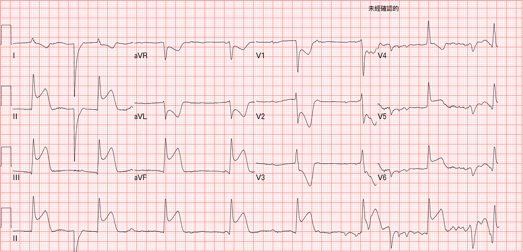
The arrival ECG is the image at the top of this post
First, let's read the ECG:
Rate: 48, SR with 3rd degree AVB, normal axis, junctional escape rhythm, STE over Inf.lead with reciprocal change over I/aVL, Lateral leads STE, STD over V1~V3
You have to consider Inf.-Post-lateral wall STEMI
First, how do you identify 3rd degree AVB?

To identify an AV block, look at the relationship between the P and the QRS.
P and QRS drift farther and farther apart, then a P isn't followed by a QRS →Mobitz Type 1
P and QRS at a fixed distance, then a P isn't followed by a QRS →Mobitz Type 2
No regular relationship in the distance between P and QRS →Complete heart block (this Case)(Fig.1)
Tips: When you see lots of P waves (more than one P between QRS and QRS), think of two situations first: AV block or blocked APCs
Next, how do you diagnose Post.wall MI?
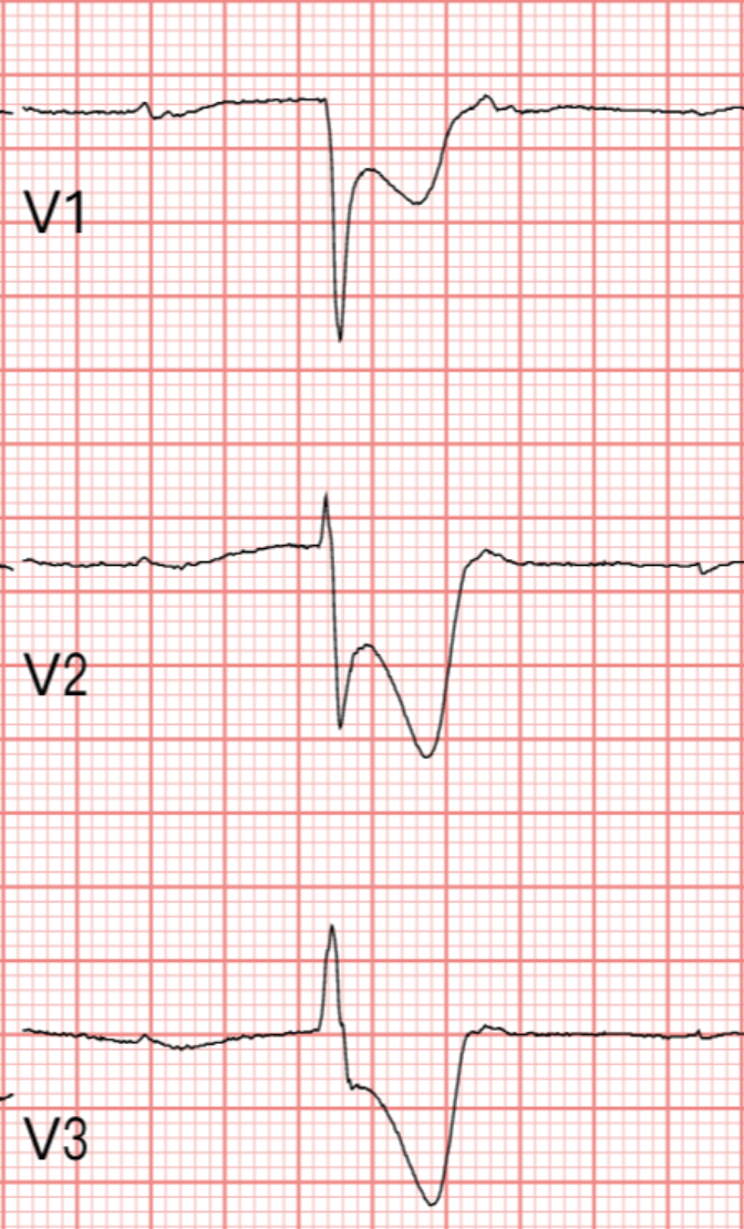
Generally, diagnosing Post.wall MI takes three criteria: (Fig.2 is V1~V3 from this Case)
1.R/S>1 in V1 or V2 (i.e., Tall RV1) 2.STD in V1~V3 3.Upright T wave in V1~V3
As I mentioned in this post, points one and three aren't actually required. Why?
Point one shows up when a Q wave has formed in the Post.wall, which corresponds to a Tall R wave in V1~V3
Point three shows up when the Post.wall MI has developed reperfusion T waves (Biphasic TWI or Deeply TWI), which corresponds to an Upright T wave in V1~V3
So if the Post.MI is in progress and no Q wave or reperfusion has appeared yet, of course all you'll see is STD in V1~V3.
The Smith ECG Blog has stressed many times: if the Maximal STD is in V2~V4, you must consider Post.wall OMI first.
Why do a Right side ECG in Inf.wall STEMI?
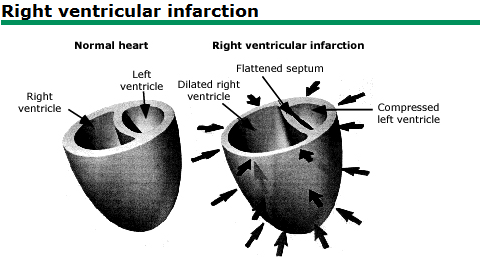
First you need to know why you'd do a Right side ECG at all. A Right side ECG tells you whether there's an associated RVMI. If there's RVMI, in management we won't give drugs that lower preload (such as NTG). Because giving something like NTG keeps fluid in the periphery, so less blood returns to the heart; and because of the RVMI, the RV dilates and the septum pushes into the LV, reducing cardiac output. Less blood back to the RV means less blood to the LV, so cardiac output drops even more, and the patient may go into shock and hypotension. (Fig.3)
Basics: About 30~50% of Inf.OMI comes with RVMI
Does NTG really drop the blood pressure?
Podcast 128 - Who Gets A Right-Sided ECG? w/ Dr. Stephen Smith & Tom Bouthillet
In this Podcast, Dr. Smith talks with a fellow prehospital EMT. In the discussion, they describe how prehospital providers worry that in inf.wall STEMI with associated RVMI, giving NTG will make the patient drop their blood pressure. The episode mentions a paper1:
The study found that in patients with Inf.wall STEMI and Non-inf.wall STEMI given NTG, there wasn't much difference between the two groups in whether it lowered blood pressure.
The paper found that after NTG, SBP dropped by ≧30 mmHg➡23.4% in inf.STEMI vs. 23.9% in non-inf.STEMI
So what does this study tell us: that, per the theory, NTG makes blood pressure crash and hurts cardiac output? Or that NTG drops blood pressure anyway, to begin with?
How can you tell there's RVMI without doing a Right side ECG?
Read the original ECG (ECG clues), using the following:
- STD in V2, combined with any of the following (this is the point emphasized more in Amal Mattu's teaching):
- STE in V1 →highly specific for RVMI
- ST isoelectric in V1
STE in V1 (Smith stresses that even 0.5 mm of STE should make you consider RVMI)
STE III >>> II (much greater than, as stressed in Amal's ECG teaching)
STE in V1>V2 (or in other words, a Flattened V2)
How to explain it➡Because on a normal ECG, V2/V3 usually have some degree of STE, so normally the ST in V2 should be higher than in V1. But if the ST in V2 is lower than in V1, that may be because of an associated RVMI, which lifts the ST in V1, so the ST in V2 ends up relatively flat compared with V1) (Note: because there may be an associated posterior OMI, V1 in RVMI won't always show STE)
- STE in aVR in the setting of Inf.wall MI➡also hints at possible RVMI, so doing a Right side ECG is a good idea, especially in every Inf.wall MI (From Smith ECG2)
How to explain it➡aVR also counts as a rightward lead on the ECG, so it can also look at the RV
After all that, which vessel was occluded in this Case?

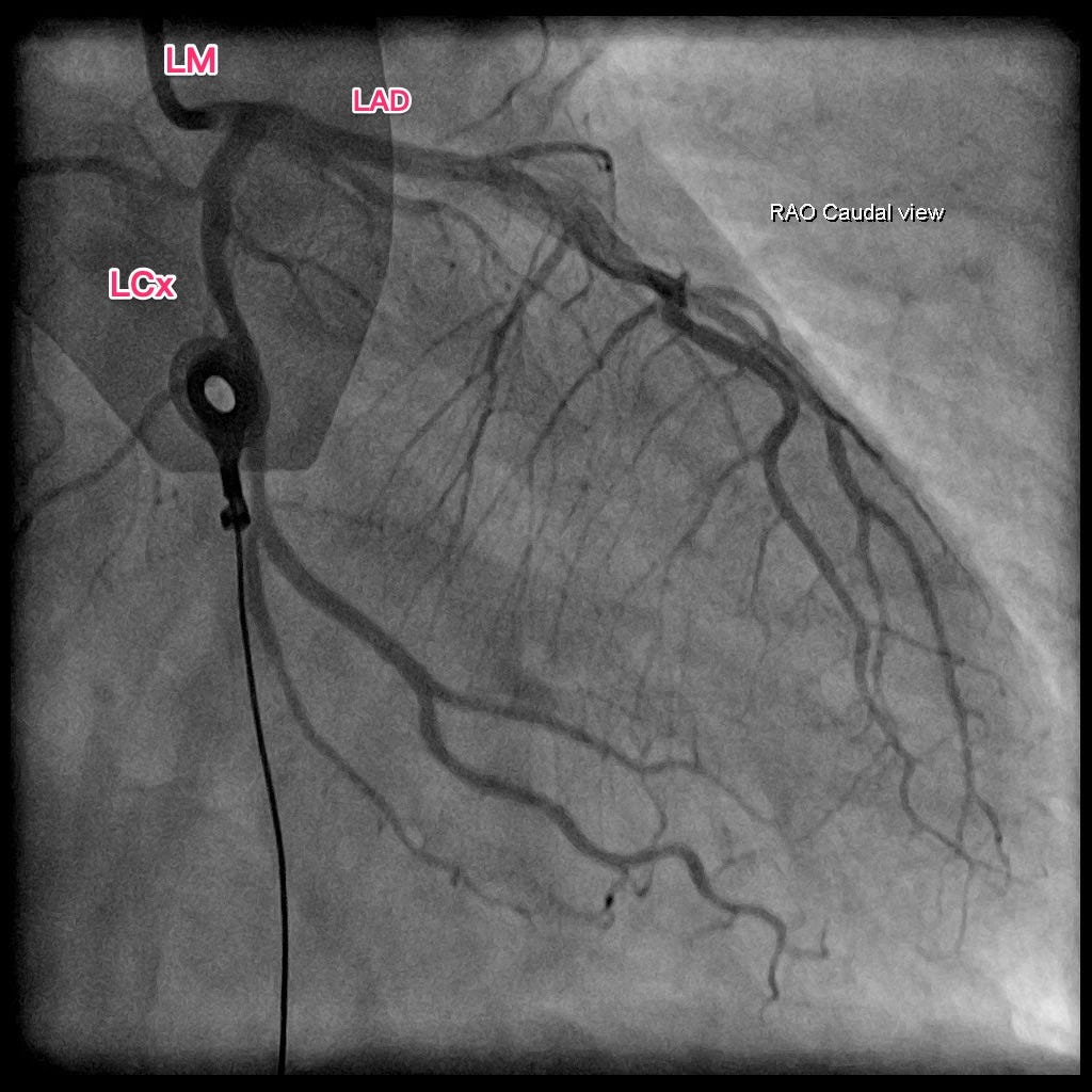
Above is the CAG (Fig.4). All three vessels wide open, as patent as can be~~~
CAG report: CAG: Insignificant CAD with marked coronary spasm over LAD-M and RCA-D
Amazing~~~
Here let's look at a figure from a paper3 (Fig.5), where it's more obvious. In the lower panel, after nitrate was given into the coronary artery, the occluded spot in the LAD opened up.

Coronary artery spasm is also called Prinzmetal (vasospastic) angina: vasoconstriction causes ischemia, producing a STEMI ECG pattern.
Here I'll mention an ECG challenge published in Circulation in July this year4. The key points:
- If the Vasospasm is brief, it can be asymptomatic but still produce the ECG changes of myocardial ischemia
- Typical angina comes from stable CAD with exertion and is relieved by rest. But that's not what happens in coronary artery spasm➡it typically occurs at rest, especially at night or in the early morning
- Arrhythmias associated with Coronary artery spasm include SB, sinus arrest, advanced AV block, Vf, asystole
- In an observation study, CAVB was the most common arrhythmia in the setting of significant CAD but no hemodynamic instability
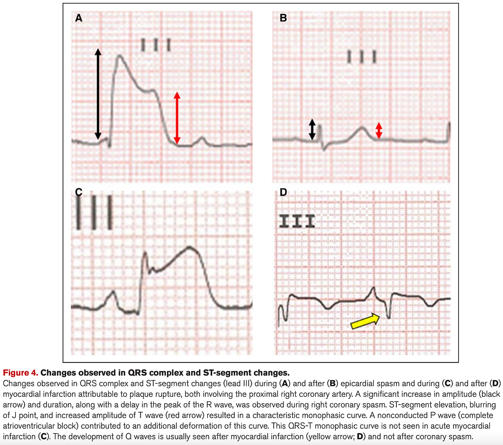
How do you tell whether STE is caused by Coronary artery spasm or by epicardial artery occlusion (AMI)? (Fig.6) →A/B are Coronary a. spasm, C/D are AMI
- STE accompanied by an increase in R wave height and width (duration) in the same lead, a bit like a monophasic curve appearing at the peak (curve at the peak➡because the R wave duration gets longer)➡QRS-T monophasic curve (panel A of Fig.6)
- This QRS-T monophasic curve isn't usually seen in AMI; rather, it's caused by repeated coronary spasm occluding the proximal portion of a main coronary artery
- Also, if pathological Q waves and T wave changes appear (Terminal TWI or Deeply TWI, i.e., Wellens’ waves), that's more of a typical AMI presentation
Additional references: 1 2 3 4
Robichaud, L., Ross, D., Proulx, M.-H., Légaré, S., Vacon, C., Xue, X., & Segal, E. (2016). Prehospital Nitroglycerin Safety in Inferior ST Elevation Myocardial Infarction. __Prehospital Emergency Care__, __20__(1), 76–81. https://doi.org/10/gmsvrq ↩︎ ↩︎
Lip, G. Y. H., Ray, K. K., & Shiu, M. F. (1998). Coronary spasm in acute myocardial infarction. __Heart__, __80__(2), 197–199. https://doi.org/10/gmvd2v ↩︎ ↩︎
Real, C., Morales, T., & Viana-Tejedor, A. (2021). An Unusual Call From the Urology Ward. __Circulation__, __144__(4), 324–327. https://doi.org/10.1161/CIRCULATIONAHA.121.055185 ↩︎ ↩︎



Thoughts on this case? Leave a comment, a like, or a reaction below.
comments powered by Disqus