Translated from the original Traditional Chinese post. Read the Chinese original →
67-year-old woman, chest pain for 1~2 days, severe pain this morning
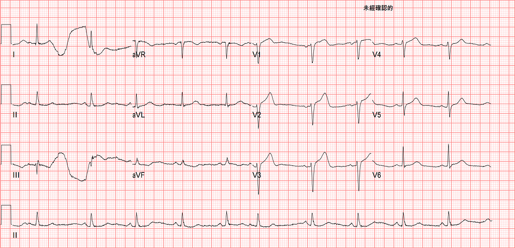
This case was from a night shift last month, in the last half hour of the morning before I was about to go off duty. Out of nowhere I was told a chest pain patient would be arriving shortly (a VIP, apparently — the patient hadn't arrived yet, but the phone call asking for a favor already had).
The nurse said, it's all on you now~~~
I thought, I've got half an hour left, what exactly do you want me to do LOL
Enough rambling, let's see what we can learn from this case!!!
When the patient arrived, she looked like she was in real pain, her whole face scrunched up from it. She said she'd had chest pain coming and going for the past 1~2 days. This morning it got severe, radiating to her back, bad enough to break out in a cold sweat.
First, let's read the ECG
Rate: 63 bpm
Rhythm: SR with APC
Axis: Normal axis
Interval: No QT prolong
Ischemia: read it using QRSTU
- No obvious pathologic Q wave
- PRWP(+)
- STE in V1~V4
- Hyperacute T wave over V2~V4
For ischemia, let's zoom in on V1~V4

V1 has 1 mm of STE
V2 has 2 mm of STE
V3 has 1.5 mm of STE
V4 has 1 mm of STE
By the definition in the STEMI criteria, across all the leads, V2~V3 in a woman of any age needs ≥1.5 mm to count as significant elevation; in the other 10 leads, 1 mm counts as elevated. Elevation in two contiguous leads is significant.
So this case absolutely, genuinely meets STEMI criteria.
Add the Hyperacute T waves in V2~V4, plus PRWP, and you can actually already diagnose LAD occlusion.
But will this ECG get the CV man to agree and activate the cath lab? More on that in a bit…….
Besides the ischemic ECG changes, the ECG in Fig.1 has an interesting artifact.


Look at the limb leads on the left first: the ECG artifact shows up in Lead I and Lead III, while Lead II has no artifact.
Then look at the diagram on the right side of Fig.2 and you can tell which lead is the problem. The left arm (LA) moved while the ECG was being recorded, so Lead I/III, which both connect to the left arm, are both affected.
If you sketch out the diagram on the right side of Fig.2, you can quickly figure out which limb is causing the ECG artifact in the limb leads. Takes 5 seconds.
Case continue
As I was saying, will subtle precordial leads STE, PRWP, and Hyperacute T waves like these convince the CV man that this chest pain is an AMI?
Every CV man has their own ruler in their head for cath lab activation, and how long or short that ruler is, we in the ED can't change.
The patient is in our hands in the ED (at least in the ED, we're the duty VS). So what's our goal?
Over the past few years, given the ECG's many qualities — it's repeatable, it's convenient — I've become deeply convinced it's an excellent tool for helping diagnose AMI. So if we have the tool, what's our goal?
The goal is to identify OMI (Occlusion MI) patients early and convince the CV man to give OMI patients reperfusion therapy. Open the vessel, and save the myocardial cells that are still left.
If the CV man sees this ECG, agrees the vessel is occluded, and activates the cath lab right away. I respect~~~
If the CV man sees this ECG and doesn't think the vessel is occluded, I also respect~~~
Medically speaking, whether it's the ED or the CV man, we're all trying to do right by the patient.
If we can't get the cath lab activated right away, that means we don't yet have enough MI evidence to convince the other side. That's my own view.
So the reason I've been trying to make my ECG reading more precise over the past few years is that I want enough evidence when I read an ECG to show that this patient needs the cath lab now, not to be parked while we trend cardiac enzymes.
There I go again with a pile of rambling from the heart.
Anyway, the CV man felt the arrival ECG (Fig.1) didn't look like an MI.
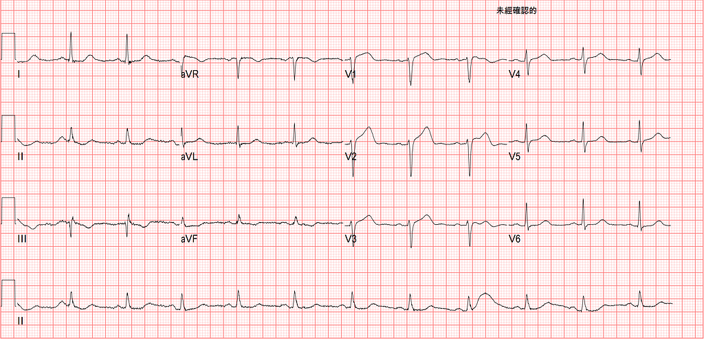
Fig.3 is the ECG 10 minutes later; the chest pain was still ongoing, but somewhat relieved. Compare Fig.3 and Fig.1 lead by lead: the Hyperacute T waves in V3~V4 have improved, and the STE in V2~V4 has come down. We call this kind of ECG change dynamic STTC, and it suggests there's a problem with the vessel.
We did an echo on the patient at the time, so let's see what findings the echo showed.
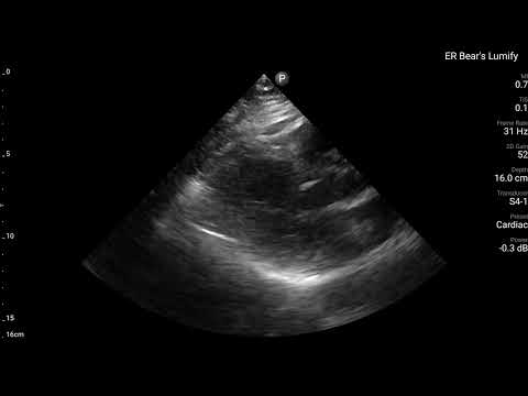
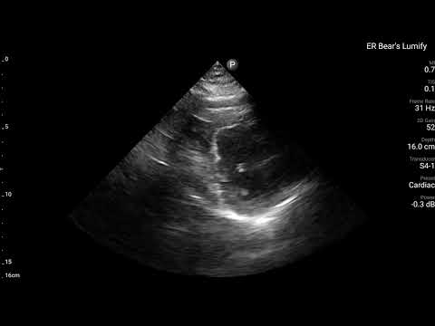
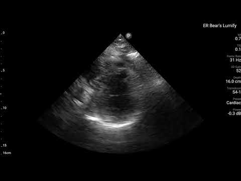
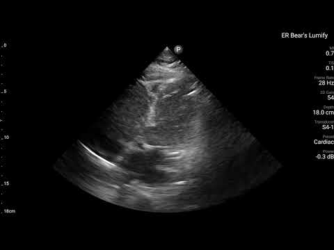
On the echo, we can see the Ant. wall supplied by the LAD is barely moving. (Best taken together with Fig.4)

The ECG shows Subtle STE/PRWP/HATW in the Precordial leads over LAD territory, plus dynamic STTC 10 minutes later, plus RWMA of the ant. wall on echo. Any way you slice it, this evidence plainly says the LAD has a problem.
Surely now we go to the cath lab????
Nope~~~~~~Orz
Then I went off shift. I told the higher-up at the patient's side that this looked very likely to be a myocardial infarction.
When I came back on shift that night, I asked the higher-up who'd been with this patient why on earth the CV man ended up doing the cath after all.
She said after I left the ED, the patient's chest pain got so bad she wet herself, and she asked the CV man what to do?! The CV man said, if it hurts that much, let's go in!!!!
I've often brought up that the 2020 ESC NSTEMI guideline says if a patient is classified as NSTE-ACS (NSTEMI+UA) and has a very high risk feature, they need the cath lab immediately, within <2 hr.
Let's review it again: look at Very High risk in the lower left corner of Fig.5.
One of the items is Recurrent/refractory chest pain
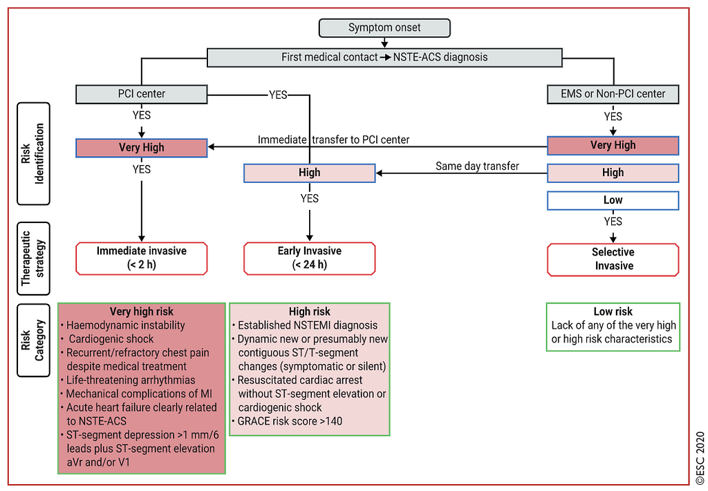
Dr.Smith has pointed out many times on his ECG blog that not just in the US but around the world, few places actually follow this Guideline and take Very high risk patients to the cath lab immediately (< 2hr) — meaning the Guideline is often violated.
That matches our own experience in the ED: the patient clearly has ongoing chest pain, but because the ECG shows no STE, we just keep trending cardiac enzymes (ongoing chest pain, with no ECG changes needed at all, already meets the indication for cath). I've also seen patients with chest pain plus cardiogenic shock where the ICU started a vasopressor and kept observing, with no cath (ACS with cardiogenic shock is an indication for cath).
Again, every CV man has a somewhat different idea in their head of what meets the indication for cath. When I think it should be done and CV thinks so too, I'm genuinely happy for the patient.
Cath report: LAD total occlusion s/p emergent PCI, Dx with NSTEMI
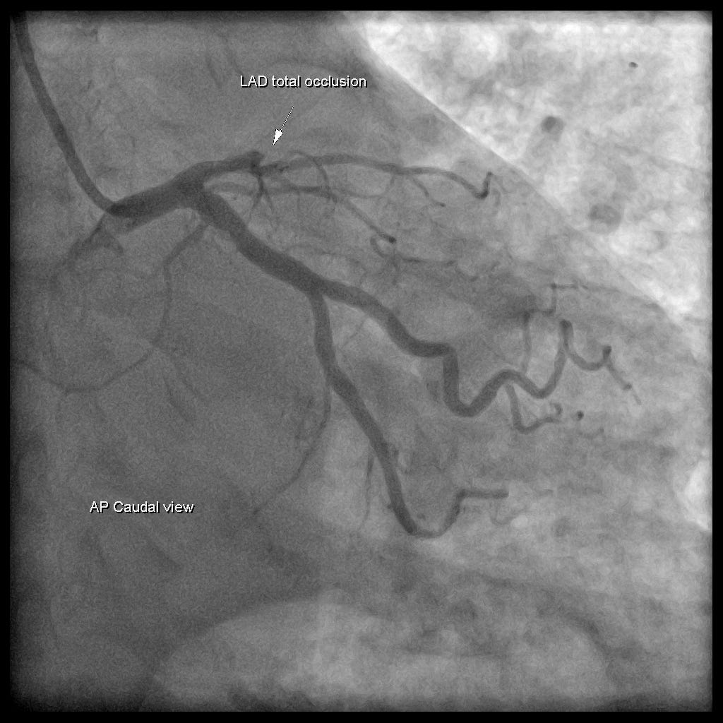
Also, because this patient had subtle STE over precordial leads, we can apply the Smith 4 variable formula to see whether LAD occlusion is likely.
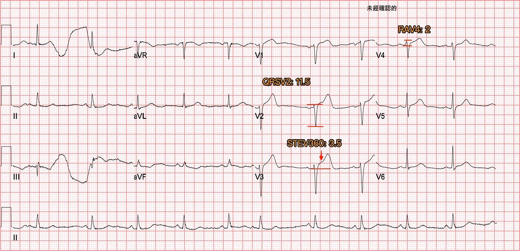
Smith 4 variable formula:formula link here
QRSV2: 11.5
- Measure the QRS amplitude in V2
STEV360: 3.5
- This value is the height above baseline 60 ms after the J point in V3 → i.e., 1.5 small boxes after it
RAV4: 2
- This value is the R wave amplitude in V4
QTc:444 ms
- Just plug the computer-calculated QTc from the 12 lead ECG into the formula
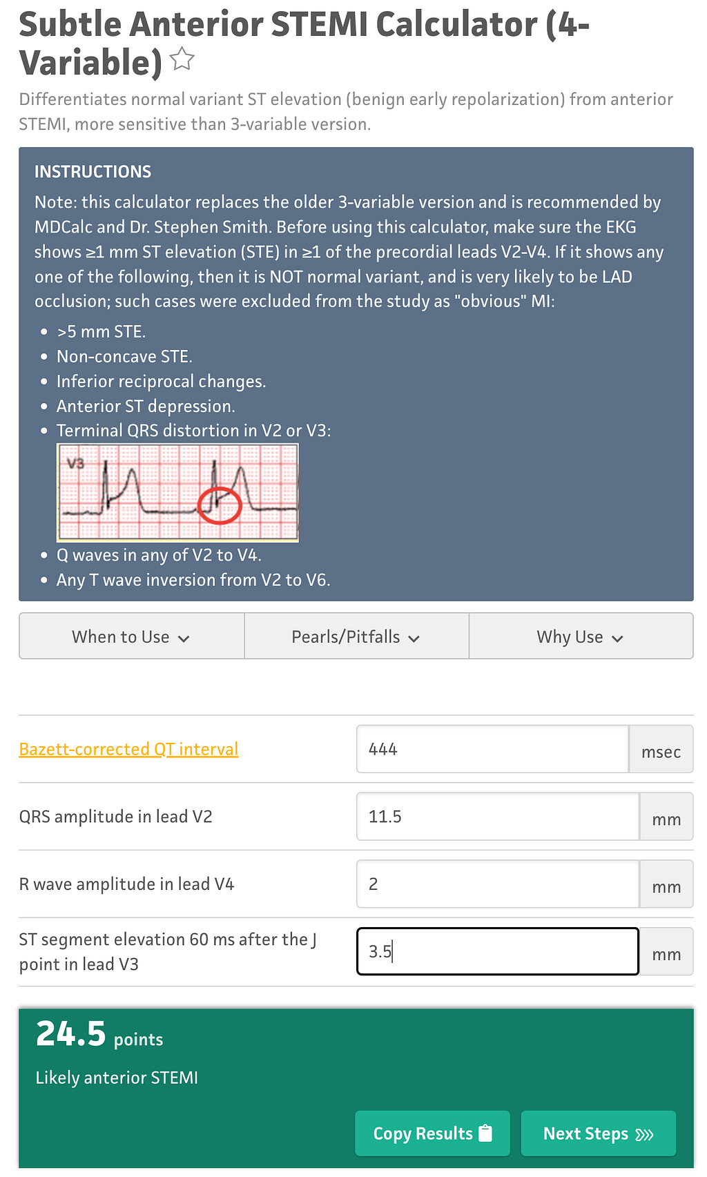
Plug the numbers into the online formula, and the result is 24.5
As long as the formula's result is >18.2, it shows likely anterior STEMI
- 83.3% sensitivity、87.7% specificity、and 85.9% diagnostic accuracy
Is this hard to calculate → not really. As long as you know where each value is measured, you can get the values you need from V2, V3, and V4 in about 1 minute.
Learning points:
- You can read ischemia using QRSTU
- Using the diagram on the right side of Fig.2, you can quickly find where a limb artifact problem is coming from
- Subtle LAD occlusion sometimes meets STEMI criteria, yet still isn't considered a problem (this is real-world reality, not an exception)
- What do we do when the arrival ECG can't convince the CV man to do a cath? We still have echo, we can still do serial ECG, and we can check whether the patient meets the Very high risk criteria in the 2020 ESC NSTEMI guideline (cath needed within <2 hr) → all of these give us more MI evidence and backup for getting the cath done
- How to tell whether subtle precordial leads STE might be LAD occlusion → you can apply the Smith 4 variable formula



Thoughts on this case? Leave a comment, a like, or a reaction below.
comments powered by Disqus