Translated from the original Traditional Chinese post. Read the Chinese original →

Narrow QRS tachycardia
50-year-old man with prior Aortic valve surgery. Chief complaint: palpitations (the ER ECG is the top image of this blog post).
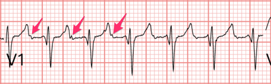
Quick read: Rate about 150, there seems to be a P, but it doesn't look like a typical P wave shape. RAD. Narrow QRS tachycardia, with obvious aVR STE with multiple leads STD
Always remember: the best lead for checking whether there's a P wave is either lead II or V1. Amal Mattu especially likes V1.
Also note that this obvious aVR STE with multiple leads STD is usually not caused by an LM, Proximal LAD, or TVD problem (see Fig.2). It's an ECG sign of cardiac ischemia caused by rate-dependent oxygen/demand mismatch. It tends to show up whenever the heart rate is too fast. Not just in PSVT — you can see it in AfRVR too. As long as the heart rate is fast, the heart beating too fast may cause transient ischemia and produce this Pattern.
So could it really be fast and also really be an AMI?
It's possible. Amal and Smith have emphasized a few key points on their blogs; let me pull them together.
Key point one: AMI rarely causes a fast heart rate. Unless there's cardiogenic shock.
Key point two: if you really do run into a fast heart rate plus chest pain/cold sweats/low blood pressure, you really can't rule out an MI coexisting with the fast rate. Of course, we've covered this situation in ACLS teaching too. This is unstable tachycardia — get the rate under control first. If after rate control this ECG pattern is still there and the chief complaint persists, then you do need to consider the DDx of the ACS sick P’t (Fig.2).
Key point three: when you see an ECG pattern like this, you must match it to the patient's clinical context. Because there are many more situations (Non-ACS sick P’t) that show this pattern.
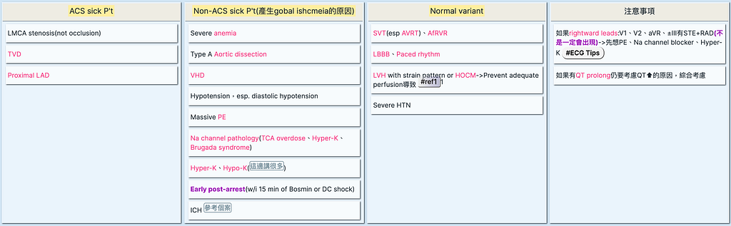
The DDx for Narrow QRS tachycardia includes the following:
- Sinus tachycardia
- AFL 2:1 or 1:1
- PSVT
Sinus tachycardia → of course, if you see obvious P waves that are upright in I/aVF/II and inverted in aVR, you can call it sinus tachycardia. So how fast can sinus tachycardia go? Maximal rate is 220-age
PSVT → if you don't see obvious P waves, you can classify it as PSVT. It can be AVNRT or Orthodromic AVRT; either way the ECG pattern will be a narrow QRS tachycardia
AFL → when we clinically see a tachycardia at 150 bpm ± 20, we have to consider AFL with 2:1 conduction. One concept here: we should say 2:1 conduction, not 2:1 block. Why? Because the AVN (AV node) here is just doing its job — its job is to keep too high an atrial rate from conducting down — so it's "conduction", letting one out of every 2 (2:1 conduction) or 3 (3:1 conduction) P waves through to the ventricle. If the AVN crashes and can't conduct, leading to a very slow ventricular rate (one P doesn't get through, the next P doesn't get through, and only the one after that gets through), that situation is called "block"; for example, 3:1 or higher is called High grade block (≥ 3:1 AVB) (Fig.3), not 3:1 conduction. And of course it's not called AFL with 3:1 block either (Flutter waves aren't that slow). This was a concept my brain somehow got tangled up on back then, so I emailed Amal Mattu, and this is the concept he wrote back to me.

I've drifted off topic a bit. Back on track~~~
Besides AFL 2:1, AFL with 1:1 conduction also needs to be considered. Of course, in that situation the rate is definitely not 150 bpm ± 20 but >200 bpm. Flutter waves run about 250~350 bpm (that's the atrial rate), so when they conduct down 1:1, the ventricle may not be able to handle it and may go into Vf (ventricular fibrillation).
Here's an example of AFL with 1:1 conduction (Fig.4), from the Smith ECG blog, describing a patient whose regular medications were Flecainide and diltiazem to control an arrhythmia.
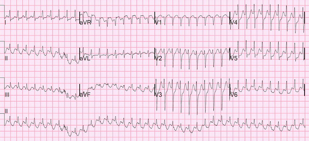
One thing to note here, which quite a few ECG blogs have mentioned: when we use a Class I antiarrhythmic to control AFL (to maintain sinus rhythm), we must add an AVN blocker. Why? Because the Class I antiarrhythmic slows the flutter waves, but once they're slow enough, the AVN lets them through 1:1 (still very fast at that point). If there's no AVN blocker to reinforce the AVN so it can block 1:1 conduction, and everything comes through 1:1, Vf can easily follow.
Tip1: To check for P waves, look at V1 or II
Tip2: DDx with aVR STE with multiple leads STD
Tips3: DDx of Narrow QRS tachycardia
Tips4: When to call it conduction, when to call it block
Tips5: If you use a Class I agent to control AFL, you must pair it with an AVN blocker



Thoughts on this case? Leave a comment, a like, or a reaction below.
comments powered by Disqus