Translated from the original Traditional Chinese post. Read the Chinese original →
So what is this twisted, distorted ECG?
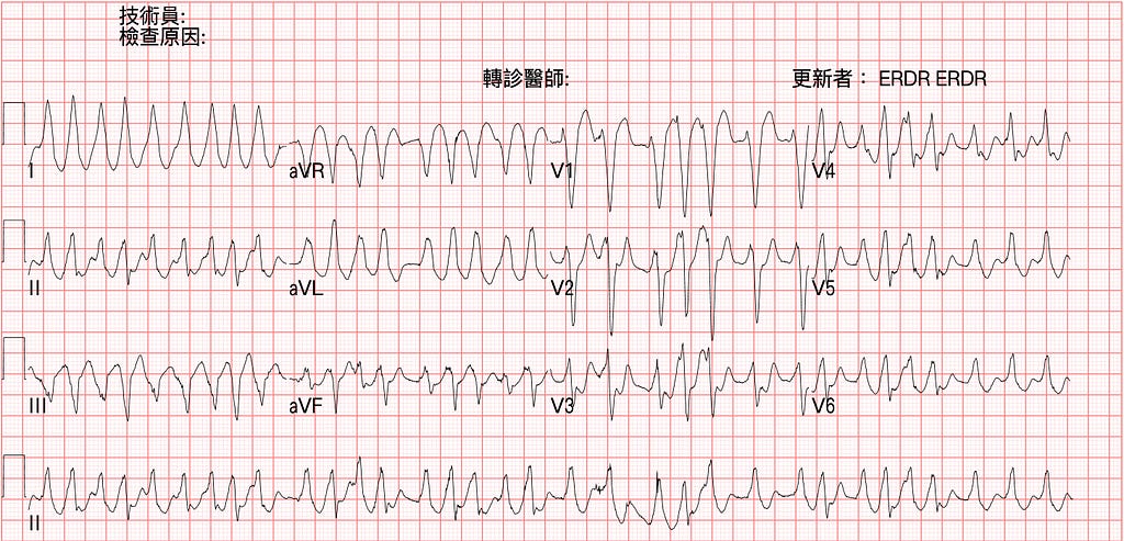
A man in his 50s went to a local hospital with palpitations and chest tightness. ECG above. The doctor on shift read it as newly onset LBBB and suspected ACS, so he was transferred to our hospital.
On arrival he was alert, BP 70/40 mmHg.
Never mind what rhythm this is for now — whether you look at the 2015 or 2020 ACLS guideline(Fig.1), it's spelled out clearly: unstable tachycardia should get Synchronized cardioversion.
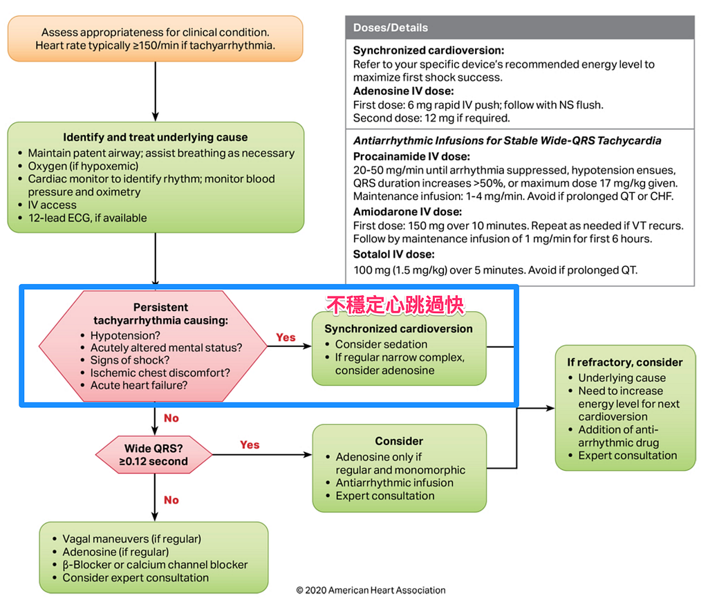
In ACLS teaching, we often use the mnemonic altered mental status, pain, dyspnea, shock, hypotension to decide whether a patient with a fast heart rate (>150 bpm) needs emergency treatment
- Altered consciousness → possibly from poor cerebral perfusion caused by the fast rate)
- Chest pain → possibly ischemic chest pain caused by the fast rate
- Dyspnea → possibly acute heart failure caused by the fast rate
- Shock → symptoms of poor peripheral perfusion: diaphoresis, cold extremities, decreased urine output, hypotension, etc.
- Hypotension
Case continue
So what rhythm is this? Fast or not? Regular or not?

Pull out the long lead II (Fig.2): very fast, some beat-to-beat rates over 300 bpm, no regularity, wide QRS, and the QRS shape keeps changing. That irregularly irregular feature means Af.
So let's start asking a few questions
🙋♂️Q1: What are the possibilities for an irregular, wide-QRS tachycardia?
- Af+BBB
- Af+WPW
- Polymorphic VT
🙋♂️Q2: How do you tell apart the DDx in Q1?
- First, what is Af+BBB (bundle branch block)?
It means Af + LBBB or RBBB. The patient already has RBBB or LBBB, so the QRS is already wide because of the BBB; add Af on top and you get an irregularly irregular rhythm with a wide QRS.
- Then what is Af+WPW?
It means there is an accessory pathway (AP from here on). After the atrium fires, the impulse doesn't take the normal SA node → AV node route. It goes down the AP instead.
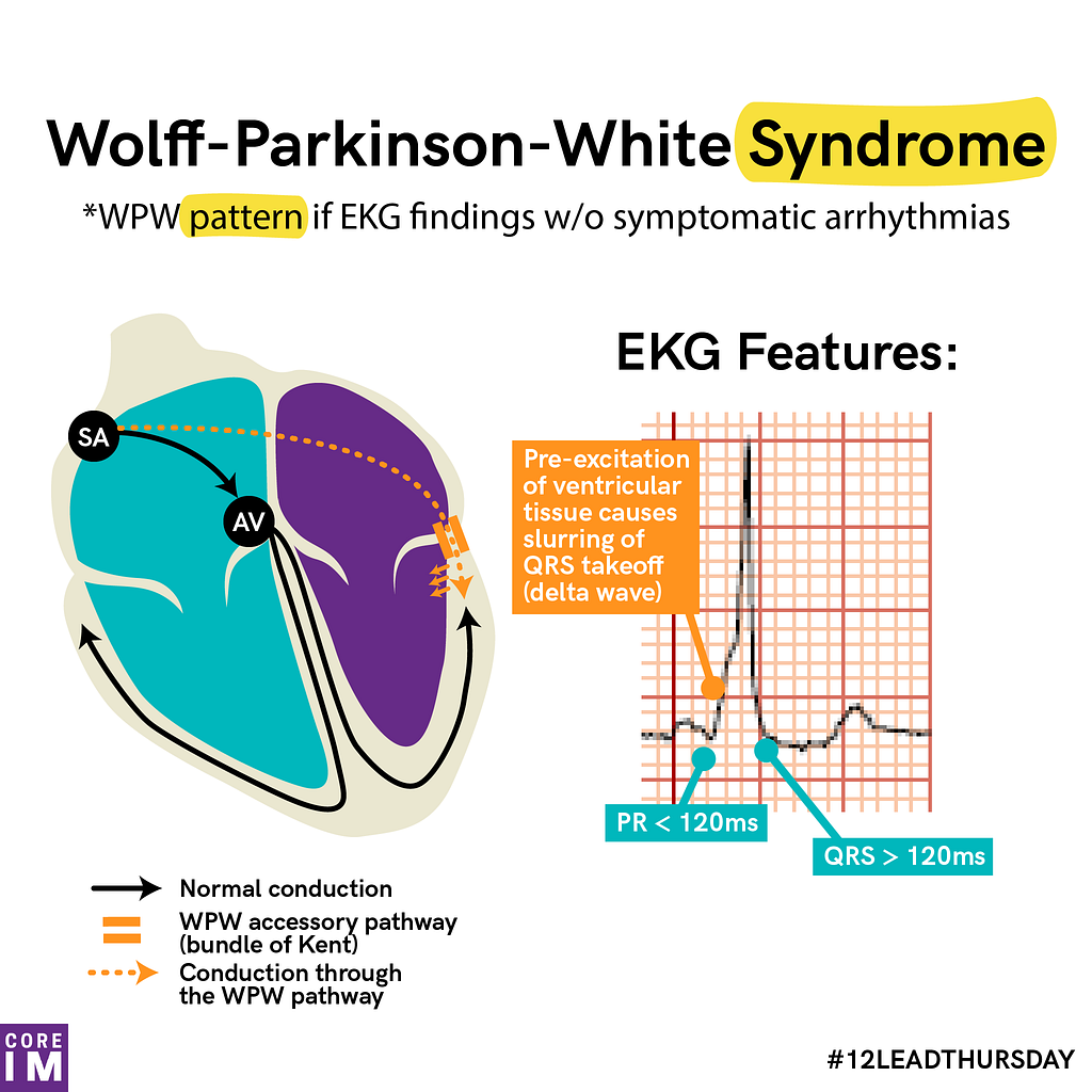
So if we see the ECG features on the right side of Fig.3 (wide QRS, delta wave, short PR interval), can we call this patient WPW syndrome?
Nope!!!!!
Having a WPW pattern ECG and having WPW syndrome are not the same thing. WPW syndrome means the patient has a WPW pattern ECG plus a symptomatic arrhythmia (follow-up question: Q3). Without a symptomatic arrhythmia, the most you can say is that this patient's ECG is a WPW pattern ECG, and send them to CV OPD to evaluate whether ablation is needed to head off future trouble.
- Next, what is Polymorphic VT?
Polymorphic VT comes in two types:
1️⃣ Type one is Generic polymorphic VT
Normal QTc
Common causes are ACS, Brugada syndrome (less common, but it belongs on the DDx)
2️⃣ Type two is Torsades polymorphic VT (TdP polymorphic VT)
The ECG before the polymorphic VT shows QT prolongation
Tips: Clinically, if QTc > 500ms, go look hard for the cause of the QT prolongation
TdP polymorphic VT also comes in two kinds:
🅰️: Acquired: caused by drugs, Hypo-K, Hypo-Mg, Hypo-Ca
- Usually QTc>600 ms
- Usually a VPC lands, causing R on T, and then TdP starts
🅱️: Congenital: caused by Congenital Long QT syndrome
- Rarely causes unstable polymorphic VT
- Preventive treatment includes Beta blockers (beta stimulation would promote TdP)
Tips: Acquired type TdP polymorphic VT is treated with beta stimulation (Overdrive pacing, isoproterenol) to speed up the heart rate and shorten the QT, and Beta blockers are contraindicated. But Congenital type TdP polymorphic VT is treated with a beta blocker
But Dr. Smith notes in this post that whether using beta stimulation agents in Congenital type TdP polymorphic VT actually makes things worse is hard to say for now1.
So can you tell from the ECG whether it's Generic polymorphic VT or Torsades polymorphic VT?
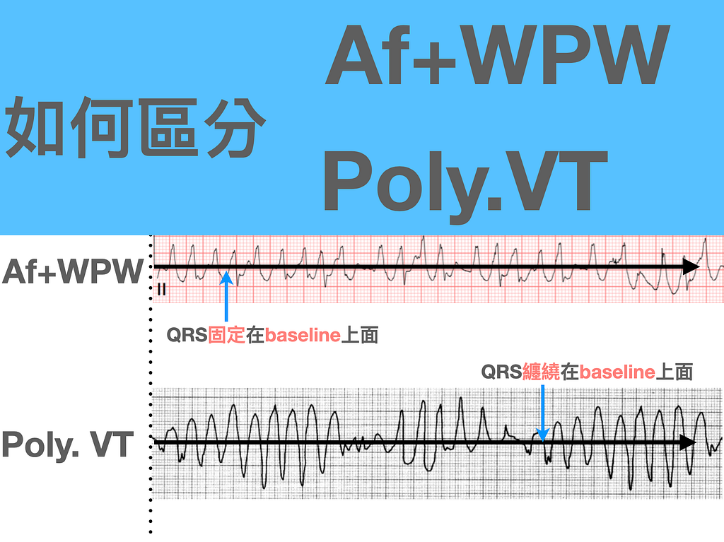
Sadly, no!!! Both of them twist around the baseline; the only way to tell them apart is to look at a prior baseline ECG for QT prolongation.
Pretty complicated, right? To simplify as much as I can, I made the mental-model diagram below (Fig.4).
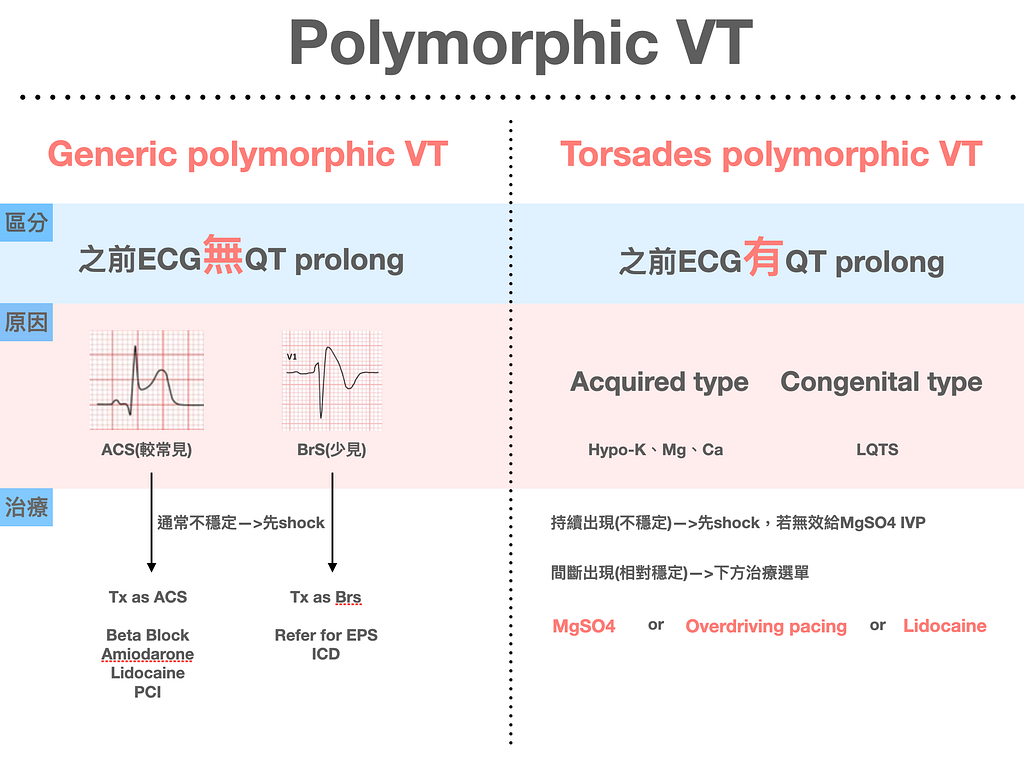
TdP polymorphic VT is a huge topic. The treatments in the figure are what's used acutely. How to give them? I'll write a separate blog post later and explain it in detail.
After all that, I still haven't gotten to how to tell Af+WPW, Af+BBB, and Polymorphic VT apart?! (This author talks way too much 😅)
Let's see where these three ECGs actually differ (Fig.5)
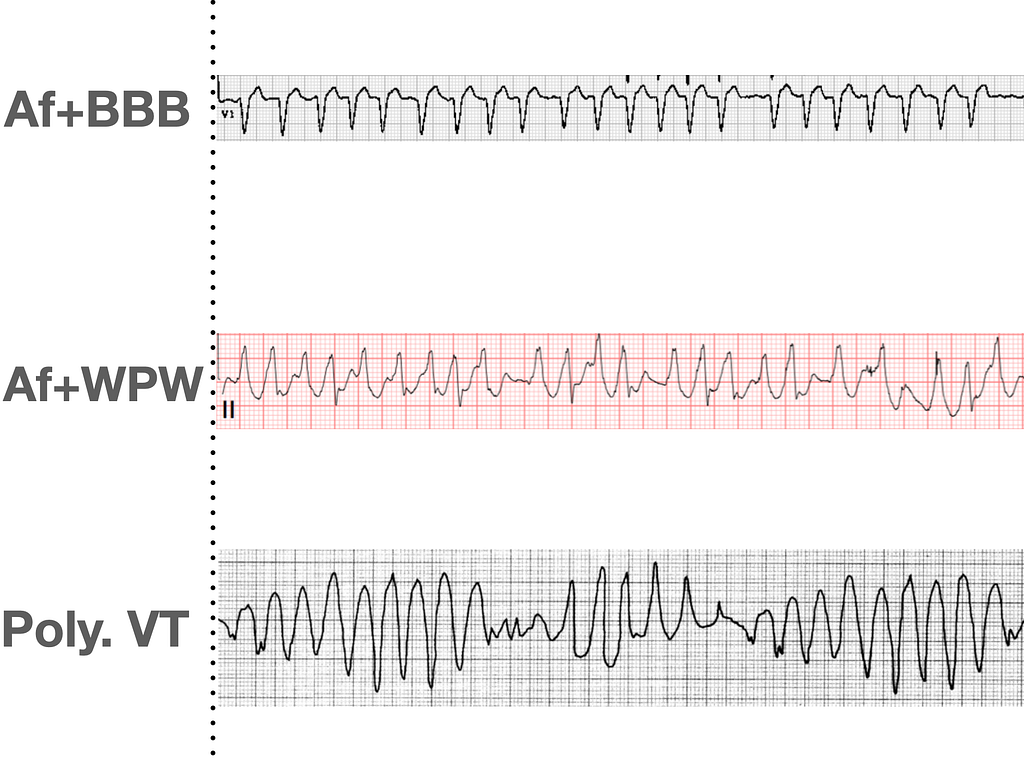
All three of these ECG morphologies are irregular, and all have wide QRS. You can separate them like this (Fig.6)
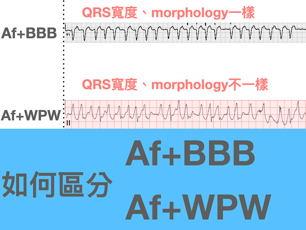
Start with the left side of Fig.6: why are Af+BBB and Af+WPW split this way?
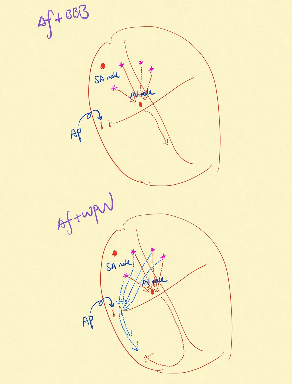
To answer that, first think about how each one conducts (Fig.7)
Af+BBB takes the normal conduction pathway (Atrial impulse->AV node)
Af+WPW takes the AP pathway (Atrial impulse->AP)
On the normal pathway, a normally functioning AV node blocks most of the atrial impulses coming down (longer refractory period). With many atrial impulses coming down (Af atrial rate: 350~600 bpm), the AV node holds its post and lets only one of those many impulses through to the ventricle.
But if there's an AP, the defining feature of an AP is a very short refractory period. So when one atrial impulse goes down the AP and another arrives right behind it, the short refractory period can't stop it. Different atrial impulses can go down hand in hand within a very short time, so the QRS morphology (ventricular depolarizations from different atrial impulses fused together) is different every beat.
Then on the right side of Fig.6, why are Af+WPW and Polymorphic VT split this way?
In one of Amal Mattu's courses, someone asked how to tell Af+WPW from PMVT (polymorphic VT) on the ECG. Amal's answer: look at whether there is a twisting baseline
I recommend Amal Mattu's EMC2 course. Two full days packed with ECG. Probably more than 400~500 tracings of reading practice. It's been running since a year ago, once every 3~4 months, and you get a certificate of completion. Of course, it's not just about collecting a boring certificate. The point is that it's a really practical ECG course. 👉 A course this good — why wouldn't you take it?
🙋♂️Q3: What arrhythmias occur with WPW?
With that background in place, let's look at what tachyarrhythmias occur with WPW.
There are only two forms of tachyarrhythmia → which is what WPW syndrome means (WPW pattern ECG + symptomatic arrhythmia)
1️⃣ The first is conduction straight down the AP (no reentry circuit) → Af or AFL
- Af or AFL, either one is a disaster!!! Why? Because the atrial rate is too fast; if it all comes down the AP, the ventricle can't take it and then goes into Vf
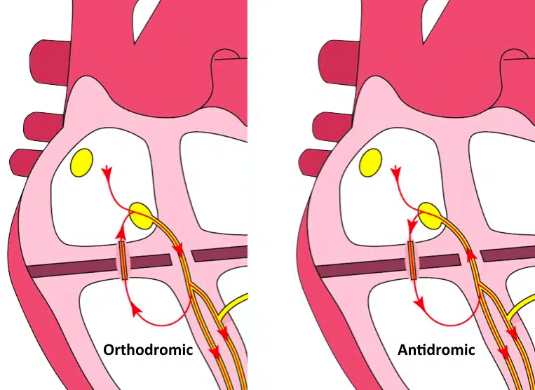
2️⃣ The second is the AP forming a reentry circuit → AVRT (a circuit between atrium and ventricle formed through the AP) (Fig.8)
AVRT comes in two kinds:
🅰️: Orthodromic AVRT: goes down the normal AV node first, then comes back up through the AP to form the loop
- From the figure (left side of Fig.8), it takes the normal pathway first, so narrow QRS (provided the baseline ECG has no BBB)
- It looks a lot like AVNRT (a loop within the AV node), but you can still tell them apart by the RP interval
- Most PSVT+WPW is orthodromic
➡️ In typical AVNRT, the retrograde P wave appears earlier, so it's either hidden inside the QRS or partially visible at the end of the QRS as a pseudo R’ wave
➡️ In AVRT, the retrograde P wave appears later, usually RP interval > 70 msec (counts as long)
Tips: Why is Typical AVNRT a short RP tachycardia, while orthodromic AVRT is a long RP tachycardia? ➡ Because AVNRT loops inside the AV node, so going up to produce the P wave takes less time / happens earlier. Orthodromic AVRT goes down, then comes back via the AP; it takes a bigger lap to complete the loop, so the retrograde P wave comes later.
- Treating Orthodromic AVRT is similar to AVNRT ➡ start with vagal maneuvers; if that fails, try adenosine and/or CCB
Tips: Watch out here → any AVN blocker can potentially trigger Af (uncommon), so when we use, say, Verapamil (a CCB), we have to observe the patient for at least 4 hours to make sure no Af develops. Otherwise, if Af develops and this patient has an accessory pathway, it becomes Af+WPW ➡ that is, if after giving adenosine/CCB or another AVN blocker the circuit is broken and only straight-down conduction remains, but you find a WPW pattern ECG, that's when to be careful. Be careful that the AVN blocker may trigger Af.
🅱️: Antidromic AVRT
- The mechanism is down the AP first, then looping back through the AV node to form the circuit, so wide QRS
- Very rare, about 5% of WPW syndrome (don't confuse it with Af+WPW: one is a tachycardia with straight-down conduction, the other is a tachycardia from a reentry loop).
- Because it goes down the AP first, the QRS is wide and it's easily mistaken for VT
- Adenosine may break this circuit, but note there is a risk of producing Af
- So in a Stable patient, use procainamide to block the accessory pathway, not an AVN blocker
🆘 Here I want to raise a question
For both Orthodromic AVRT and Antidromic AVRT, I mentioned that AVN blockers may trigger Af. So can we really give an AVN blocker (usually adenosine) before we're sure that this narrow QRS tachycardia isn't an Orthodromic AVRT using an AP, or that a regular wide QRS tachycardia isn't antidromic AVRT?
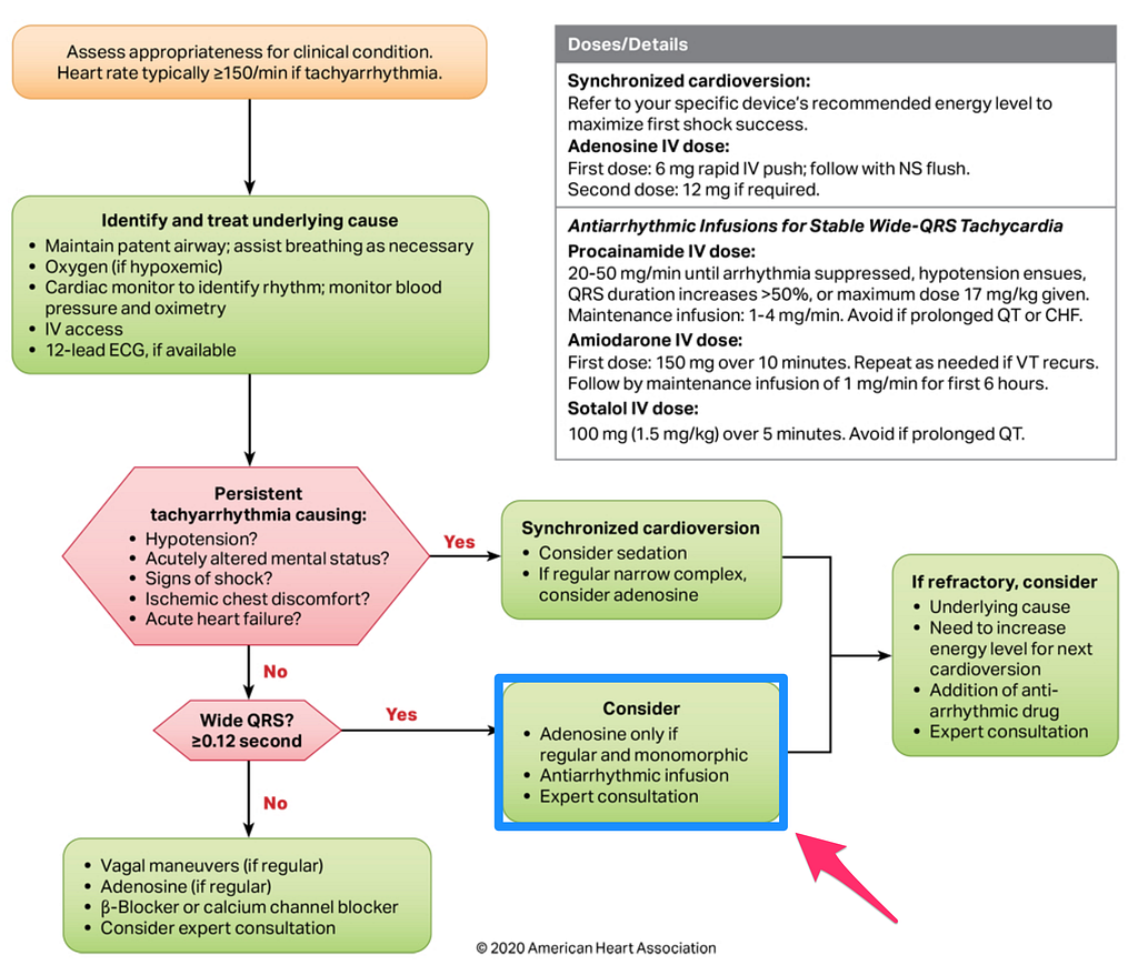
Look at the ACLS 2020 Tachycardia algorithm in Fig.9. It says plainly that for a stable, wide-QRS tachycardia, you can try adenosine (what if it's a wide QRS tachycardia from antidromic AVRT?)
This article2 mentions that adenosine can induce Af, with an incidence ranging from 1%~12% depending on the dose (I haven't run into it myself clinically).
So my own takeaway: if clinically you hit a narrow QRS tachycardia (SVT) or wide QRS tachycardia (SVT or VT?) and we break the reentry circuit with adenosine, then the repeat ECG has clues that it may be WPW with an AP — remember to observe the patient for 4 hours to make sure no adenosine-induced Af appears. Only then is it reasonable to send them home. Otherwise, if Af develops and goes down the AP, it becomes Af+WPW, which puts the patient in extreme danger.
Case continue
Because this was an unstable tachycardia (hypotension) in the ED, we gave biphasic synchronized cardioversion at 200 J. After the shock he returned to a normal rhythm, with a WPW pattern ECG morphology.
🙋♂️Q4: If you choose to shock Af+WPW, synchronized or unsynchronized?
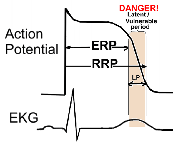
- The defibrillator fires by catching the R wave of the QRS. If we go unsynchronized, it's easy to hit the T wave and cause Vf. Only if synchronized really won't fire (the machine keeps failing to find the QRS) do you choose unsynchronized.
- There are many case reports of unsynchronized shocks landing on the wrong spot on the T wave and causing Vf, mainly through the R on T phenomenon. And these cases were almost all Af+WPW. See Fig.10: the vulnerable period is in the T wave region. So never fire there, or the heart will go haywire.
Learning Points:
- How to identify an unstable tachycardia
- How to tell apart Af+WPW, Af+BBB, PMVT
- What arrhythmias occur with WPW (WPW syndrome)?
- AVN blockers may trigger Af
- If you choose electrical therapy for Af+WPW, remember to press the sync button
Dr. Smith’s ECG Blog: Polymorphic Ventricular Tachycardia — link ↩︎ ↩︎
Chakraborty, P., & Subramanian, A. (2017). An unusual complication of adenosine administration in supraventricular tachycardia. __Indian Pacing and Electrophysiology Journal__, __18__(1), 36–38. https://doi.org/10.1016/j.ipej.2017.11.002 ↩︎ ↩︎
emDOCs.net — Emergency Medicine EducationElectrical cardioversion in the ED: who crashes and how to improve — emDOCs.net — Emergency Medicine Education — link ↩︎



Thoughts on this case? Leave a comment, a like, or a reaction below.
comments powered by Disqus