Translated from the original Traditional Chinese post. Read the Chinese original →
Answering a reader's ECG questions
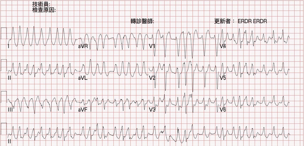

OnDecember 17 last year I published a post, and recently a reader asked some questions about it. I'll answer them here.
The questions boil down to a few points:
Q1 → Could Af+VT be happening at the same time?
Q2 → I thought WPW driven by A-fib goes down the Accessory pathway. If it takes the same path, shouldn't the QRS complexes be more stable — shouldn't the beat to beat QRS difference be smaller than this?
Q3 → I thought this EKG had AV dissociation, favor VT?
Q4 → This ECG has a very wide QRS complex, favor VT?
Q1 → Could Af+VT be happening at the same time?
Let's start with the first question. Could Af+VT occur at the same time and produce an ECG like Fig.1 Left?
For you to get Af and VT together, the atrium would need pacemakers firing all over the place, and when those impulses come down they'd also have to land right in a reentry pathway inside the ventricle. (The more common mechanism of VT is a reentry rhythm.)
But in an Af rhythm, with discharges firing chaotically from many sites in the atrium, when multiple impulses conduct downward, the firing site in the atrium isn't fixed, so the time it takes to reach the ventricle isn't fixed either. It's really hard to do that without breaking up the VT's reentry rhythm.
So can Af+VT coexist? Very hard~~~~~
What's more likely to show up is Af+junction escape rhythm or ventricular escape rhythm.
That is, there's a pacemaker beating in the atrium (Af), and also a pacemaker beating in the ventricle (JER or VER).
Let's look at an example:

On this ECG you can see fibrillation waves in V1 (Af atrial rate: 350–600 bpm).
But is this Af? Isn't Af an irregular irregular rhythm? So why are the RR intervals the same?
Because there's a Complete heart block (CHB, so the Af can't conduct down. That's why an escape rhythm arises in the ventricle → if it comes from pacemaker cells near the AV node, it's an escape junctional rhythm; if it comes from ventricular pacemaker cells further down, it's an escape ventricular rhythm)
Clinically, what could cause CHB like this? Besides conduction abnormalities after an MI, you also have to think about Digoxin overdose, which can do this too.
Digoxin is a cardiac glycoside, commonly used for rate control in Af to keep it from going too fast. So when it's taken in excess (a suicidal overdose, or levels running too high when renal function worsens)
ECG changes you can see with Digoxin use include those described in the figure below.
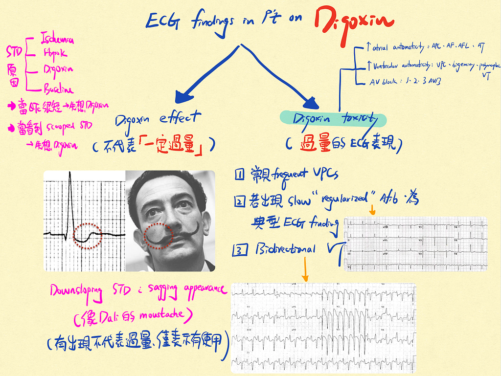
Fig.3 is a Notability note I wrote earlier. Reposting it here for another review.
Simply put, when a patient is on Digoxin, there are two kinds of ECG changes:
- Digoxin effect: digoxin effect doesn't necessarily mean overdose; it only tells you the patient is taking digoxin. Commonly Downsloping STD (Scooped STD) (note: Hypo-K can also give a similar STD)
- Digoxin toxicity: the ECG findings that show up with overdose
ECG Tips: When you see Scooped STD, think Digoxin effect first, especially when the QTc is very short
Some ECG changes you can see in Toxicity:
① Commonly frequent VPCs
② Slow "regularized" Af, if present, is the classic ECG finding (Digoxin toxicity)
③ Bidirectional VT
Also, since Digoxin is a cardiac glycoside, it ⬆︎ atrial and ventricular automaticity, which makes the following ECG changes likely:
- When ⬆︎atrial automaticity → APC, Af, AFL, AT
- When ⬆︎ventricular automaticity → VPC, bigeminy, polymorphic VT
Slow regularized Af is the classic Digoxin toxicity ECG finding. So why does it change like that?

Fig.4 explains why an Af rhythm in Digoxin toxicity becomes a regular rhythm instead of an irregular irregular rhythm.
① In Af the atrium fires chaotically all over
② Digoxin blocks conduction before the AV node
③ Pacemaker cells at the A-V junction or in the ventricle start firing (because nothing gets through from above, the lower levels fire spontaneously)
Putting all of the above together, including the extended explanation of digoxin toxicity: Af + VER or JER is the more likely possibility.
Q2 → I thought WPW driven by A-fib goes down the Accessory pathway. If it takes the same path, shouldn't the QRS complexes be more stable — shouldn't the beat to beat QRS difference be smaller than this?
The reason the QRS morphology in Af+WPW keeps changing shape comes down to the very short refractory period of the accessory pathway: it can't stop the next atrial discharge from slipping in right after the previous one. So the QRS morphologies get superimposed on each other.
Af fires at a rate of 350–600 bpm, which is very fast, and above all, each firing site in Af sits at a different distance from the accessory pathway. So the QRS morphology produced when one impulse reaches the accessory pathway and conducts into the ventricle gets superimposed on the QRS morphology produced by another firing site from the previous atrial discharge conducting down through the accessory pathway into the ventricle. The overall QRS morphology keeps changing and is never the same.
So the key point is that the accessory pathway's refractory period is too short, which is what makes the QRS morphology keep changing.
Q3 → I thought this EKG had AV dissociation, favor VT?
AV dissociation means the QRS after a sinus P wave has nothing to do with it (i.e., the P wave wasn't captured, and the following QRS is fired by the ventricle's own pacemaker, not by an impulse conducted down from above).
In an RWCT (regular wide QRS complex tachycardia), if you see AV dissociation, you do need to consider VT (and think of Complete AVB too). But Fig.1 is an irregular WCT.
Usually we'd consider the following Rhythms:
- Af+WPW
- Polymorphic VT
- Af + aberrancy conduction
And Fig.1 is Af+WPW; if you look closely, you really shouldn't be able to identify any P waves. And of course you can't assess whether a P wave was captured to produce the following QRS.
Q4 → This ECG has a very wide QRS complex, favor VT?
There are two kinds of VT:
① Monomorphic VT (when we say VT in general, this is what we mean)
- Accounts for >90% of VT
- Feature: QRS morphology and amplitude → stay constant
② Polymorphic VT
- 10% of all ventricular arrhythmia
- Feature: QRS morphology and amplitude → keep changing
In an RWCT (regular wide QRS tachycardia), can we use QRS duration to infer whether it's VT or SVT with aberrancy?
I recently read a passage on the Smith ECG Blog about very wide QRS duration ➡ in a WCT, a QRS duration > 140 ms has roughly moderate specificity favoring VT, but a duration of 160~180 ms strongly favors VT1
But if you rely on QRS duration alone to call VT, you may well get shot down.
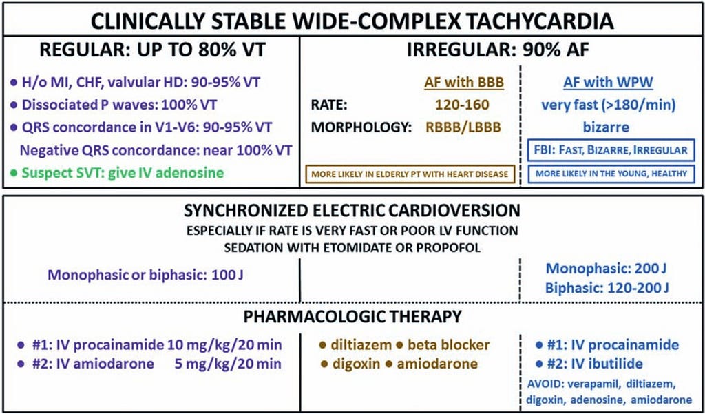
But let's look at what this 2019 AJEM article on how to approach WCT says2
- Any time you see an RWCT → over 80% are VT
- If the history includes MI, CHF, or VHD, 90–95% are VT
Hmm…… so any time you see an RWCT, if you guess VT, you'll be right 8 times out of 10 — the odds are really high (cringe attack — seems you don't even need all those VT vs. SVT with aberrancy criteria; just guess VT first and you're good😅)
The ECG master Amal Mattu also often says in his ECG teaching that he doesn't favor using all the various criteria to decide whether it's VT, for several important reasons: applying all those criteria takes too much time. And if you conclude it's SVT with aberrancy, the EP man may still go in and find it's VT (cringe attack, round two😂). That's the same as us missing VT in the ED and not treating the VT patient.
Dr. Smith’s ECG Blog: An unstable wide complex tachycardia resistant to electrical cardioversion — link ↩︎ ↩︎
Littmann, L., Olson, E. G., & Gibbs, M. A. (2019). Initial evaluation and management of wide-complex tachycardia: A simplified and practical approach. The American Journal of Emergency Medicine, 37(7), 1340–1345. https://doi.org/10.1016/j.ajem.2019.04.027 ↩︎ ↩︎



Thoughts on this case? Leave a comment, a like, or a reaction below.
comments powered by Disqus