Translated from the original Traditional Chinese post. Read the Chinese original →
With LVH, how do you tell whether there's also an AMI?
The week before last, Amal (Mattu) posted a tweet on Twitter showing everyone just how hard it is to pick out a STEMI in LVH. Flip it around, and the secondary STTC of LVH changes the ST segments and T waves in ways that make it even easier for us to mistake LVH strain pattern changes for STEMI. Lately both Amal and Smith have written quite a bit about LVH. There's only one conclusion………
LVH is so damn hard, and there are no rules!!!!!!
Is that the end of this post? Of course not~~~~~
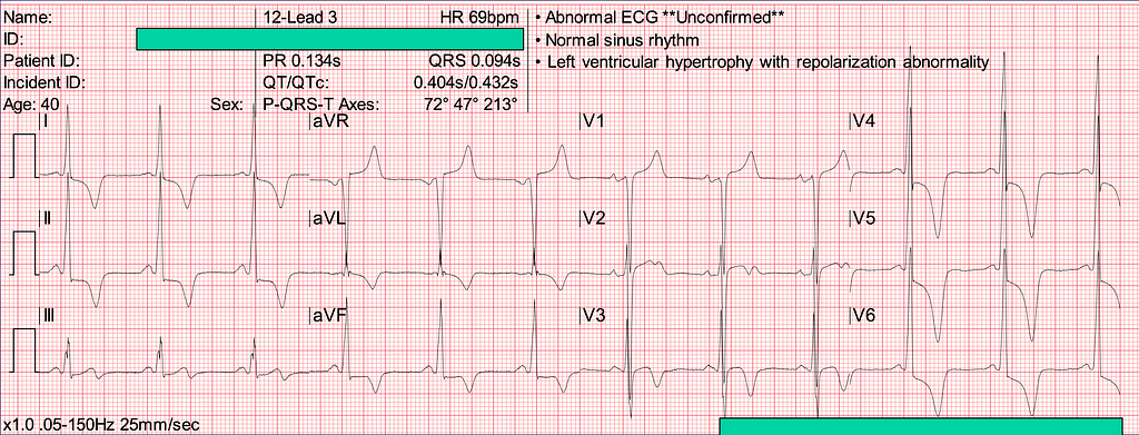
Let's look at this tweet. Try practicing on it first before you look at the answer — that's better!

If there are no rules, why bother looking? My own take: there's order in the chaos! We can't possibly Call CV to vouch for and help read the ECG every time an LVH with strain pattern has STE while we're on shift in the ED. That would really make others think we in the ED aren't professional. Of course, if you truly can't rule out AMI, you still have to consult — after all, this is "the care the patient deserves", not something as simple as the ED and cardiology playing a guessing game.
So first we need to know some general rules: what the usual LVH morphology and strain ECG pattern look like~~ Within those general rules, look for anything that's a bit different. If there is, could there possibly be an AMI? That's when it lets us raise our level of alertness for this patient.

General rules
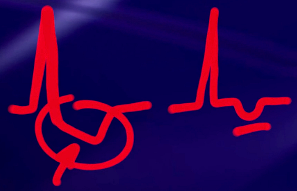
- Understand LVH with strain pattern ➡ there may be STE over R’t precordial leads with L’t precordial leads STD, I/aVL STD (I/aVL may also have STE)
- Strain type pattern ➡ you can see the STD is asymmetric; with ischemia it's more likely to be symmetric
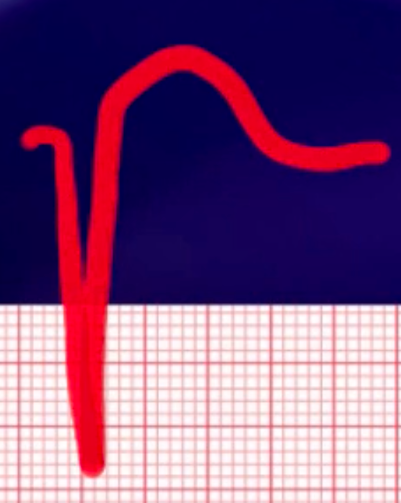
- Very deep S waves are common in the R’t precordial leads, so there will be STE (because of appropriate discordance)
- In high voltage situations, appropriate discordance will also be seen
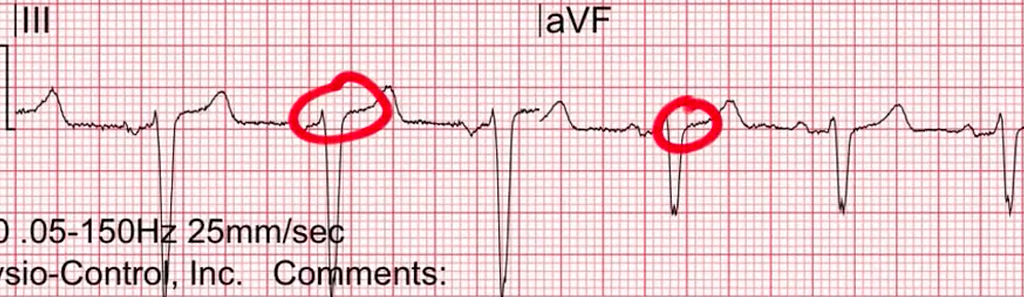
- aVR STE in LVH is something you can see, and it isn't ischemia
- LVH can also show Giant T wave syndrome
Smith also covered how to distinguish LVH with STEMI in recent posts. I've excerpted the key points below. If you're interested, check out this one and this one
Emre Aslanger's article1 describes using a value of 1/6 (about 16.6%) (to distinguish LVH vs STEMI); here you can see it's applied as STE/R-S. Stephen Smith points out something very important: STE in LVH rarely exceeds 4 mm, so if you use the STE/R-S>25% from Armstrong's article, the measure becomes very insensitive. (Sensitivity too low)
Is there any way to tell whether STE is caused by LVH or STEMI?
Some people think LVH has typical asymmetric TWI while ACS has typical symmetric TWI?
- No evidence or data support this argument
Can concave vs convex STE tell them apart?
- LVH usually has concave STE, but it can also have convex STE. The same goes for STE in ACS. So this morphology can't distinguish them either
Can the Modified Sgarbossa criteria be used in LVH?
- This refers to the Armstrong et al. article, which holds that STE in V1~V3 with STE/R-S>25% can be considered Ant. wall STEMI
- Stephen Smith thinks this article's study method wasn't appropriate. A more appropriate method would be to collect and compare serial ECGs in the leads with High voltage and STE, divide them into with/without LAD occlusion, and then look at the STE/R-S ratio. That would be more correct.
Use the T wave instead of the ST segment?
- It's currently unclear how T wave height or morphology changes in LVH under ischemia.
Can LVH use a T/QRS like the one for LV aneurysm?
- It's currently unclear whether the T/QRS>0.36 rule can be applied to ECGs of LVH patients to identify STEMI

Tips 1: For whether LVH is accompanied by STEMI, there currently aren't any good criteria. All you can do is look at lots of LVH ECGs and get yourself familiar with the ECG morphology of LVH with strain pattern
Tips 2: ECG voltage criteria alone aren't enough to diagnose LVH; they need to be accompanied by non-voltage criteria
Tips 3: In the setting of LVH, comparing with prior ECGs is very important for telling whether there's a STEMI
Tips 4: When you see an ECG pattern that leans toward OMI, you may not be able to diagnose LVH with AMI right away, but it can raise your level of alertness — do frequent Serial F/U ECGs!
Additional references: 2 3 1 4 5
Recognizing electrocardiographically subtle occlusion myocardial infarction and differentiating it from mimics: Ten steps to or away from cath lab ↩︎ ↩︎
LVH with anterior ST Elevation. When is it anterior STEMI? ↩︎
A man in his 50s with anterior ST elevation and a “tall T wave in V1” ↩︎
Amal mattu’s ECG Case of the Week — October 4, 2021 — ECG Weekly ↩︎
Amal mattu’s ECG Case of the Week– September 27, 2021 — ECG Weekly ↩︎



Thoughts on this case? Leave a comment, a like, or a reaction below.
comments powered by Disqus