Translated from the original Traditional Chinese post. Read the Chinese original →
64-year-old man with epigastric pain, chest pain, and upper back pain
This Case stuck with me, mainly because of how the patient presented.
A 64-year-old man with a history of diabetes and hyperlipidemia, who'd had three stents placed for CAD. But he hadn't been following up for the past few years.
He started feeling unwell in the morning. The pain began in the epigastrium, then spread to his chest, upper back, and jaw.
Pain bad enough to break into a cold sweat…… nauseated, wanting to vomit.
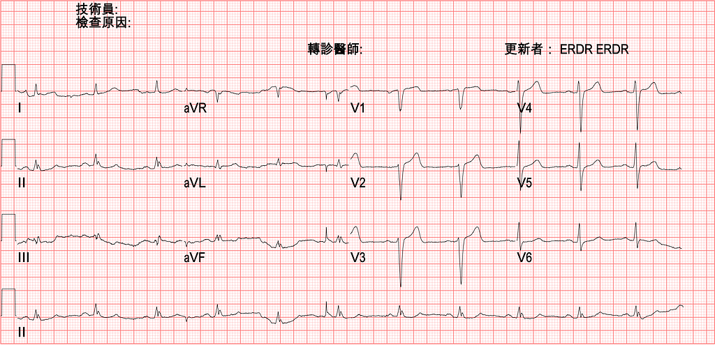
Let's first read the ECG on arrival (Fig.1) and see what clues we can pick up.
Rate →67 bpm
Rhythm →SR
Axis →Normal axis
Interval →No QT prolong (QTc: 435 ms)
Ischemia: PRWP
The baseline isn't very stable, but you can also see low voltage (Sensitive criteria with I+II+III < 15 mm).
First, a few questions to ask yourself:
Q1: Can the Smith 4 variable formula be used in this Case?
Q2: Can the T/QRS > 0.36 rule be used in this Case?
Q3: If our initial ECG is a non-diagnostic ECG, what do we do next?
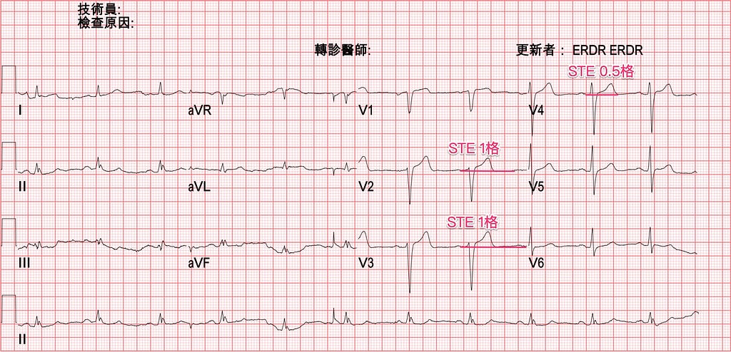
Let's answer the first question first. There's PRWP; could this be an LAD problem? This Case has 1 small box of STE in both V2 and V3 (Fig.2). Could this be a subtle STEMI due to LAD occlusion?
First of all, the Fourth Universal Definition of Myocardial Infarction in 20181 already clearly defined that, to diagnose STEMI, the criteria in V2/V3 differ from the other leads:
- Men, <40 y/o: 2.5 mm high
- Men, ≥40 y/o: 2 mm high
- Women, regardless of age: ≥1.5 mm high
So although this Case has 1 small box of STE in both V2/V3, neither meets STEMI criteria.
Next, can we use the Smith 4 variable formula to see whether there's a subtle LAD occlusion?
To use this formula, you first have to know its exclusion criteria
First, know that if there's an obvious AMI, you can't use this to calculate; it will give a serious false-negative:
5 mm STE →exclude
- Non-concave STE → if straight or convex upward STE →exclude
- Inferior reciprocal changes → if there's reciprocal change →exclude
- TQRSD seen →exclude
- Q waves in any of V2 to V4 → means AMI may already have happened →exclude
- Any T wave inversion from V2 to V6 → means possible AMI followed by reperfusion →exclude
- BBB →exclude
- LVH or LV aneurysm →exclude; false-positives may occur
Never forget: don't use the formula indiscriminately; false-negatives or false-positives can occur. Before using it, first exclude obvious AMI and everyone the article excludes, then use it.
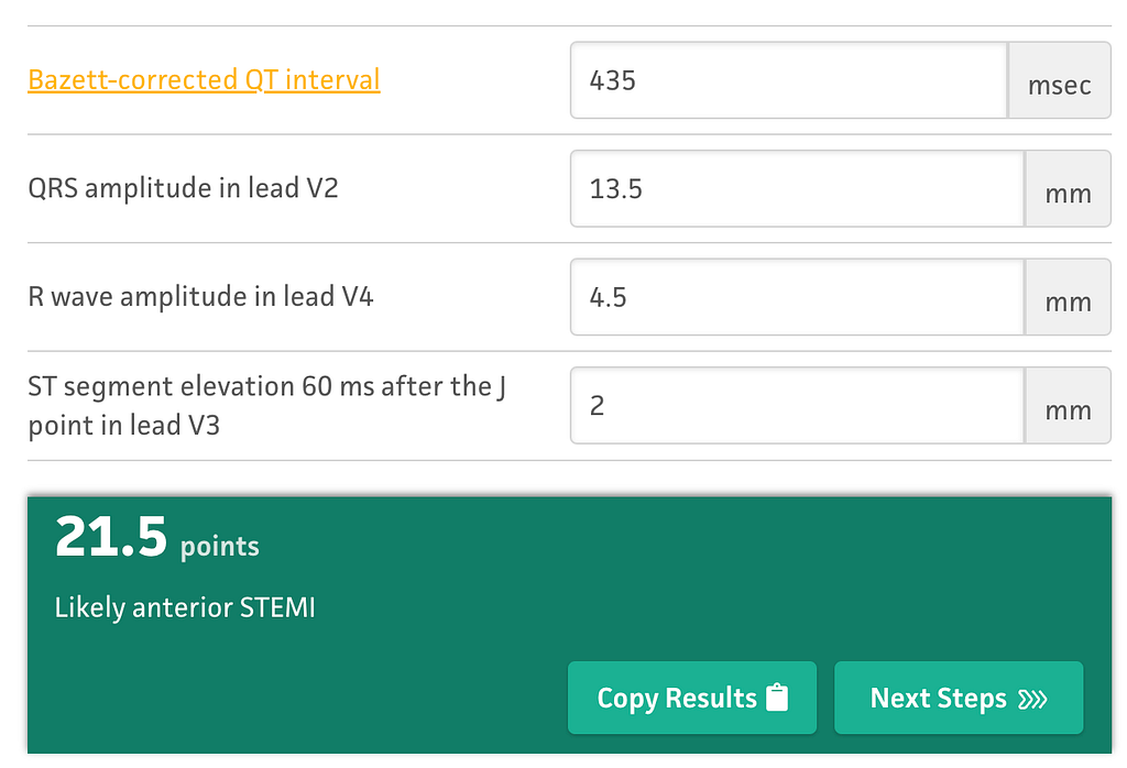
In this Case, although none of V1-V4 is a QS wave, V1 is an rS wave (r wave <1 mm). If you really want to use a formula, it should be the LV aneurysm with STE vs. STEMI one, T/QRS.
So, for patients who fit LV aneurysm with STE, can the Smith 4 variable formula be used at all?
This REBEL EM Blog article says2:
One HUGE CAVEAT, is that this equation should not be used in patients with LVH or LV aneurysms as this can cause false positives.
In plain words: the Smith 4 variable formula should not be used when the ECG has LVH morphology or an LV aneurysm with STE pattern; it may lead to false positives.
If we force the formula anyway, it comes out to 21.5 →Likely anterior STEMI (but it may be a false-positive)
Next, can the T/QRS > 0.36 rule be used?
To use this rule, you first have to know which situations it applies to. When we're trying to distinguish LV aneurysm with STE vs. STEMI, that's when this rule applies.
This rule can be used when at least one lead in V1-V4 shows a QS wave (meaning no R wave at all, or the r wave < 1 mm).
Applying the rule:
- If any lead in V1-V4 has a T/QRS ratio > 0.36, acute STEMI is the more likely diagnosis, though subacute STEMI is also possible
Applied to this Case (Fig.1), you can see that no lead has a T/QRS ratio > 0.36.
But…… and it's exactly this "but"
If this equation is used with chest pain lasting more than 6 hours, false-negatives can happen. That is, there's an AMI, but the occlusion has been there a while, so the T wave isn't as tall anymore. Looking at it as a T/QRS ratio, the numerator gets smaller, so the chance of reaching > 0.36 drops.
So for chest pain over 6 hours, if you apply this rule and it isn't > 0.36, it could still be a false-negative~~~
Now for the third question. If the initial ECG is non-diagnostic for us, what else can we do?
Do an Echo?
Wait for the TnI?
Keep F/U ECGs?
What guidelines currently tell us what to do? Does calculating a Heart score help?
Let's first look at the ACS algorithm in ACLS 2020
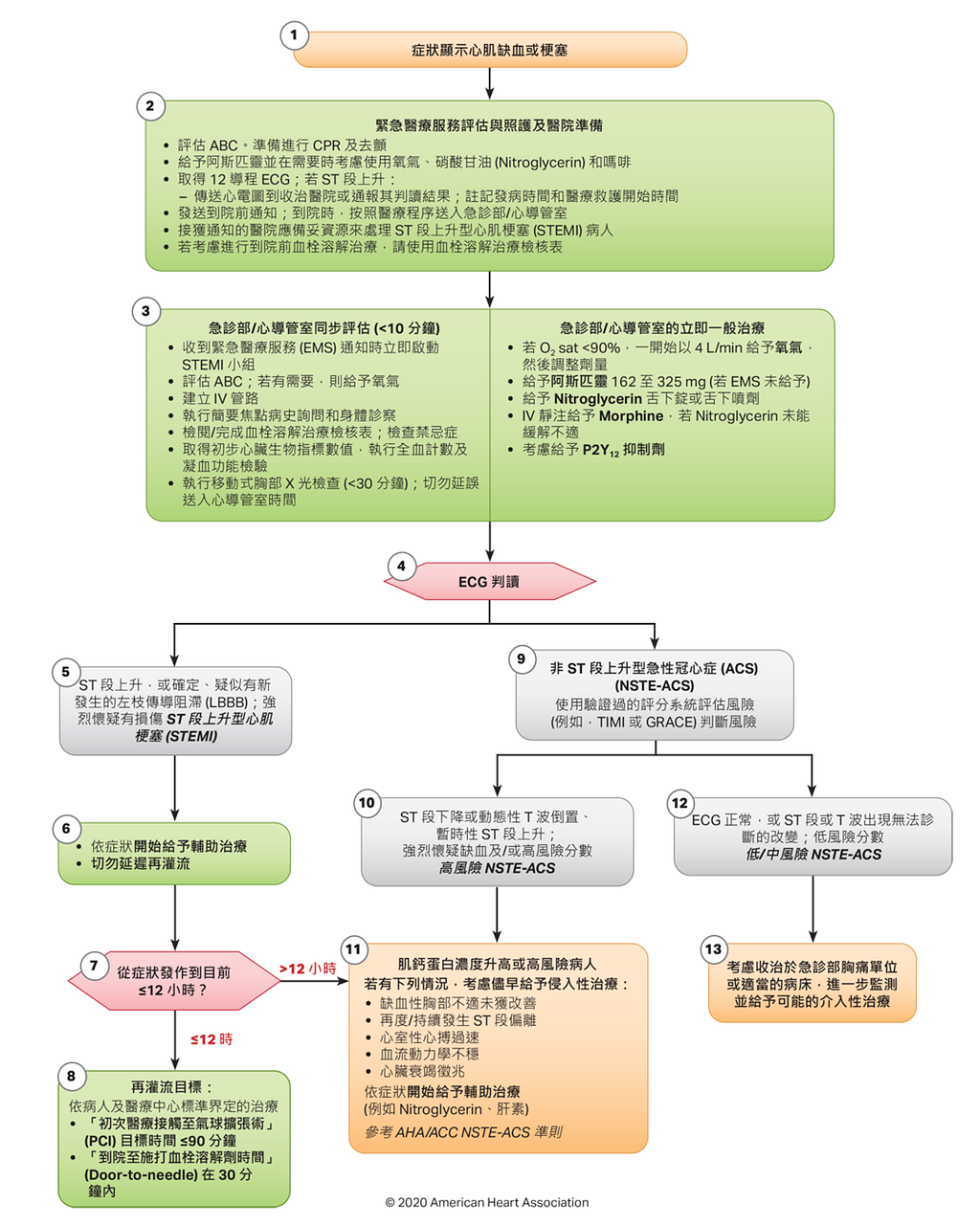
In the ACLS 2020 ACS algorithm, after the 12 lead ECG, patients are split into two big groups:
The first group is STEMI/Newly onset LBBB
The second group is NSTE-ACS (UA+NSTEMI is now called NSTE-ACS)
First, there's one fact we need to know.
newly onset LBBB ≠ STEMI
In the past, new or presumably new LBBB with AMI symptoms used to be considered a class I indication for reperfusion therapy. But the 2013 ACC/AHA STEMI Guideline removed this3.
The relevant passage is here:
New or presumably new LBBB has been considered a STEMI equivalent. Most cases of LBBB at time of presentation, however, are “not known to be old” because of prior electrocardiogram (ECG) is not available for comparison. New or presumably new LBBB at presentation occurs infrequently, may interfere with ST-elevation analysis, and should not be considered diagnostic of acute myocardial infarction (MI) in isolation
What it means is that most patients arriving at the hospital don't have an old ECG to compare with. New/presumably new LBBB is uncommon and shouldn't be used on its own to diagnose AMI.
Also, another article4 notes that, in fact, among patients presenting to the ED with ACS symptoms, the probability of AMI doesn't differ much whether they have new LBBB, old LBBB, or no LBBB: 7.3%, 5.2%, and 6.1% respectively.
So if you send everyone with LBBB to the cath lab, how high is the false-positive rate?
Pretty much sky-high XD
I'm getting sidetracked…….
In Fig.3, if it's judged to be NSTE-ACS, it has to be further split into high-risk NSTE-ACS and low/intermediate-risk N STE-ACS
High-risk NSTE-ACS presents on ECG as follows (Fig.3-step 10):
- STD > 0.5 mm
- Dynamic STTC/TWI during pain or discomfort (reperfusion rhythm appears)
- transient STEMI (<20 mins, STE >0.5 mm)
Low/intermediate-risk NSTE-ACS presents on ECG as follows (Fig.3-step 12):
- ST deviation < 0.5 mm
- TWI < 2 mm
- Normal ECG
Splitting into these two groups ties into the direction of subsequent management and treatment.
When the ECG shows high-risk NSTE-ACS, and Troponin(+) or high risk features are present → consider early invasive treatment (meaning cath as early as possible) (step 11 in Fig.3)
When the ECG shows low/intermediate-risk NSTE-ACS, consider admitting to a Chest pain unit (CPU).
Come on~~come on~~come on
A bed in Area A observation, that's our CPU (I think most hospitals don't have a dedicated chest pain observation area). Back when I was still in residency, at my home training hospital, Taipei Veterans General Hospital, there was a chest pain clinic. Pretty much every chest pain was yours to see; I just don't know whether it still exists.
So at step 13 in Fig.3, i.e. once in the CPU, what needs to be done?
The ACLS 2020 text says to do the following:
- Serial ECG
- Keep trending biochemical markers of myocardial injury (Troponin, CK-MB)
- Arrange advanced non-invasive cardiac imaging:
- Transthoracic echocardiography (2D Transthoracic Heart Echography )
- Myocardial perfusion nuclear imaging (Myocardial Perfusion Scintigraphy: MPS)
- Computed tomography coronary angiography ( Coronary CT Angiography)
- Cardiac magnetic resonance imaging (Cardiac Magnetic Resonance)
Items 2, 3, and 4 of the advanced non-invasive cardiac imaging are really not suited to the ED, at least in most EDs in Taiwan.
So to sum up, serial F/U ECG + enzyme + POCUS are the extra things we can do when the initial ECG is a non-diagnostic ECG.
So what does last year's 2022 ACC expert consensus on managing acute chest pain in the ED8 say (Fig.4)?

When symptoms suggest ACS, first get an ECG. After that, it splits into four paths: (Fig.5)
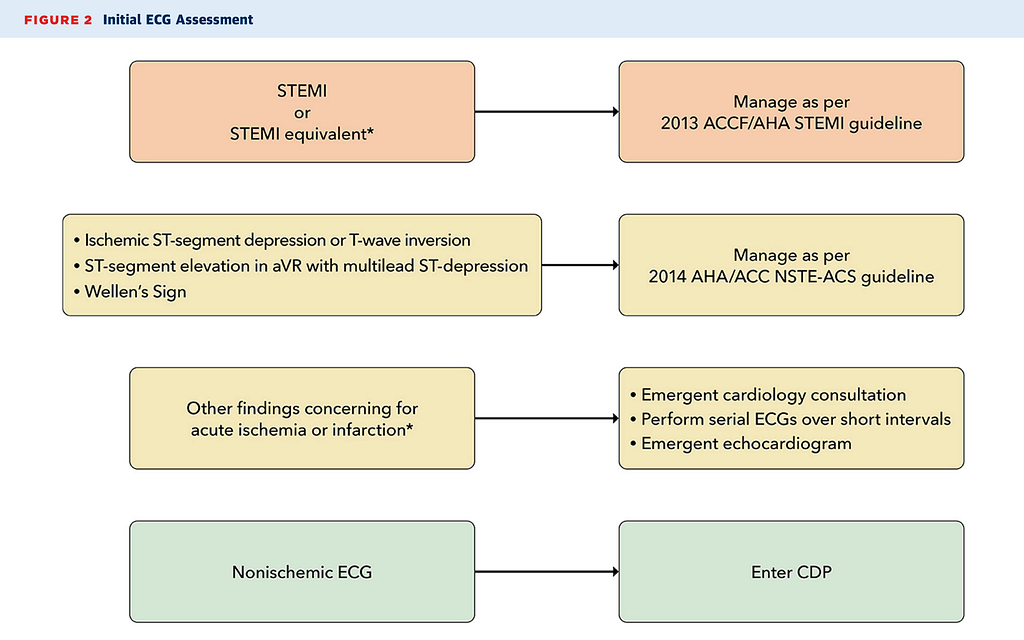
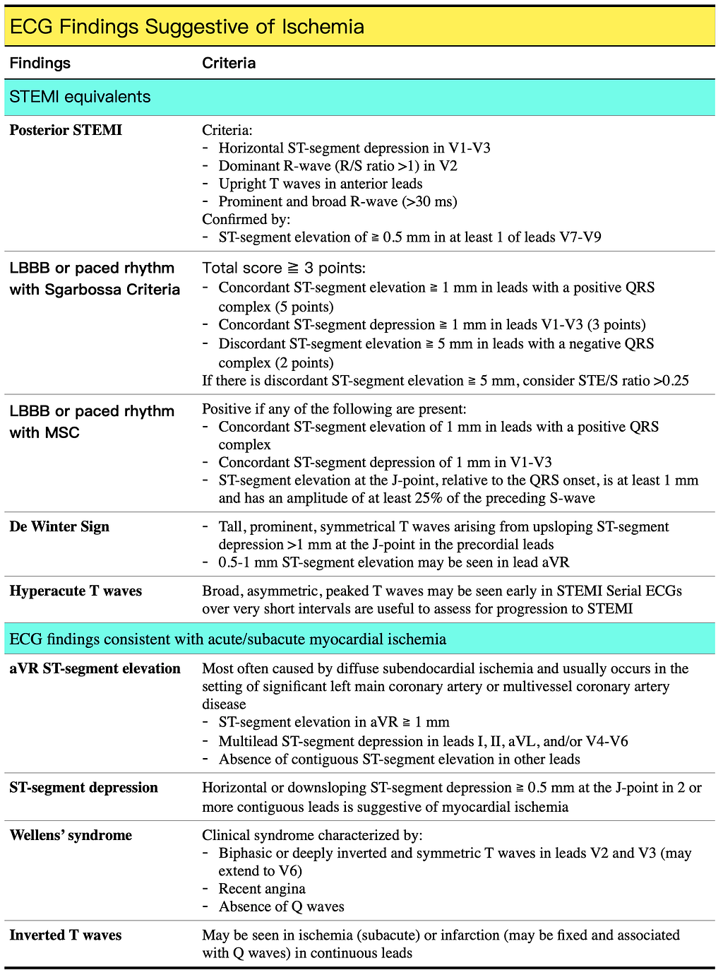
Orange: STEMI or STEMI equivalent seen (see the top half of Fig.6) → management per the 2013 ACC/AHA STEMI guideline (Bear: ESC has a 2017 STEMI guideline)
Yellow 1: ECG shows possible ischemic changes (see the bottom half of Fig.6) → management per the 2014 ACC/AHA NSTEMI-ACS guideline (Bear: ESC has a 2020 NSTEMI guideline)
Yellow 2: If the ECG changes aren't Orange or Yellow 1 but ischemic changes are still suspected → consult the CV man, Serial F/U ECG (15–20 minutes), Heart POCUS
Green: If non-diagnostic ECG → enter the CDP (clinical decision pathway)
So once it's deemed a non-diagnostic ECG, which paths can you take? (Fig.7)
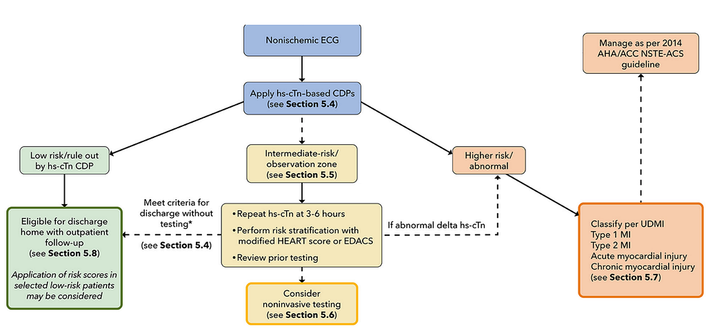
At this point, apply hs-cTn-based CDPs for risk stratification:
Low risk: can be ruled out
Intermediate risk: observe
High risk: abnormal → per the 2018 UDMI, distinguish type 1/2 MI and acute/chronic myocardial injury → manage per the 2014 ACC/AHA NSTEMI-ACS guideline
So what hs-cTn-based CDPs are there? (Fig.8)
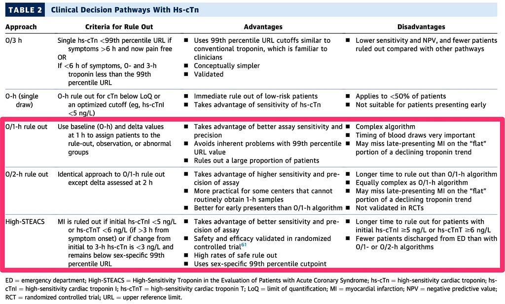
The paper8 recommends using ESC 0/1 h, ESC 0/2 h, and High-STEACS; these three are more effective (more patients ruled out) and safer (fewer patients missed) than ESC 0/3 h.
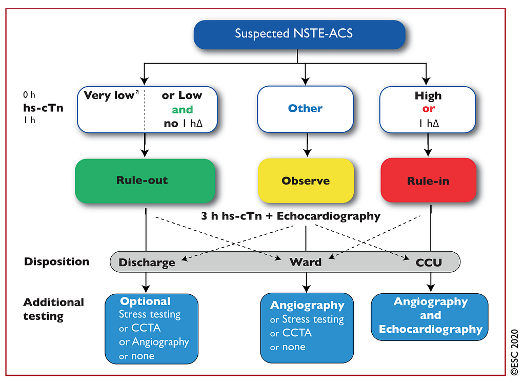
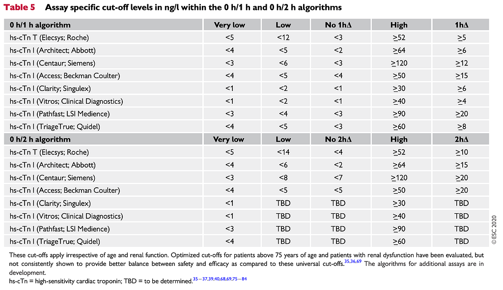
How do you apply ESC 0/1 h and ESC 0/2 h? (Read Fig.9 + Fig.10 together)
The Fig.9 flowchart comes from the ESC 2020 NSTEMI guideline. It incorporates the 0-hour rule out → meaning that if ACS symptoms ≧3 hours and the 0 h hs-cTn is Very low (see the values in the first row of Fig.10), you can rule out MI (Low risk).
- The 0-hour rule out is not for patients with symptoms < 3 hrs. Never forget this!!!!
- Remember to find out which manufacturer's hs-cTn assay your own lab uses (Fig.10)
If it's under 3 hours, the 0 h hs-cTn is low "and" the ∆ against the 1 h hs-cTn (No 1h∆ → see the values in the third row of Fig.10), meeting this condition also means you can rule out MI (Low risk).
If the 0 h hs-cTn meets High "or" 1h∆, rule in MI (High risk).
If the 0 h hs-cTn meets neither rule in MI nor rule out MI, it falls into Intermediate risk and needs observation.
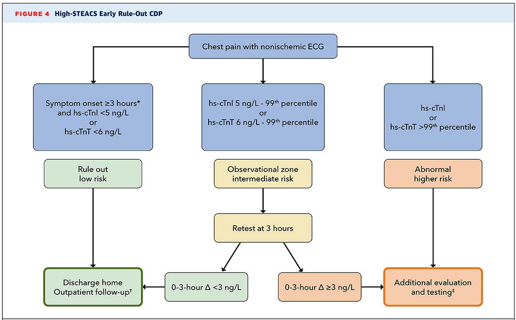
Fig.11 is another recommended CDP; if you're interested, see the figure above.
Besides Troponin measurement, which ACS patients still need risk stratification?
This paper8 describes three groups of patients who need further risk stratification:
- Patients ruled out (rule out MI) by conventional cTn
- Patients classified as rule out MI by an hs-cTn-based CDP (i.e. the low risk group)
- Patients classified into the intermediate risk group by an hs-cTn-based CDP
Quite a few hospitals still use conventional cTn (this is not hs-cTn), or use hs-cTn but don't display the value when it's below the 99th percentile of the URL → because sensitivity and NPV aren't good enough to rule out MI on their own, risk scores are recommended. Commonly used ones are the Heart score and the EDACS risk score.
The Heart score was mainly designed for risk stratification in undifferentiated chest pain, low risk. It doesn't apply to people already diagnosed with MI.
The EDACS risk score is also commonly used in low risk patients to further assess their MACE (major adverse cardiac event) (Fig.12).

Why run additional risk score rules on these patients? Mainly because a certain proportion of these patients still get missed due to false-negatives. Pairing them with these score rules lowers the chance of misjudgment. (If you're interested, see p.17-p20 of this paper8).
OK~~~ sidetracked again, back to this Case
So this Case's initial ECG was either a non-diagnostic ECG, or, possibly because the chest pain had lasted more than 6 hours, the T/QRS ratio > 0.36 rule was already giving a false-negative.
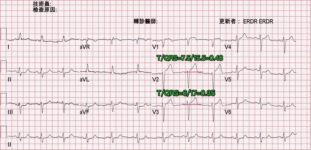
On the ECG one hour later (symptoms worse), you can see the T/QRS ratio > 0.36 rule still applies. The T wave in V2 got bigger; applying this rule, the T/QRS ratio in V2 now exceeds 0.36, favor OMI (Occlusion MI). And the inf.leads now show reciprocal STD changes, with minimal STE over aVL. This ECG pattern is very likely due to a proximal LAD occlusion.
The patient's symptoms came and went; when the pain hit, he'd pound his chest hard like a gorilla.
In the meantime we consulted the CV man; the ED consult reason was recurrent chest pain.
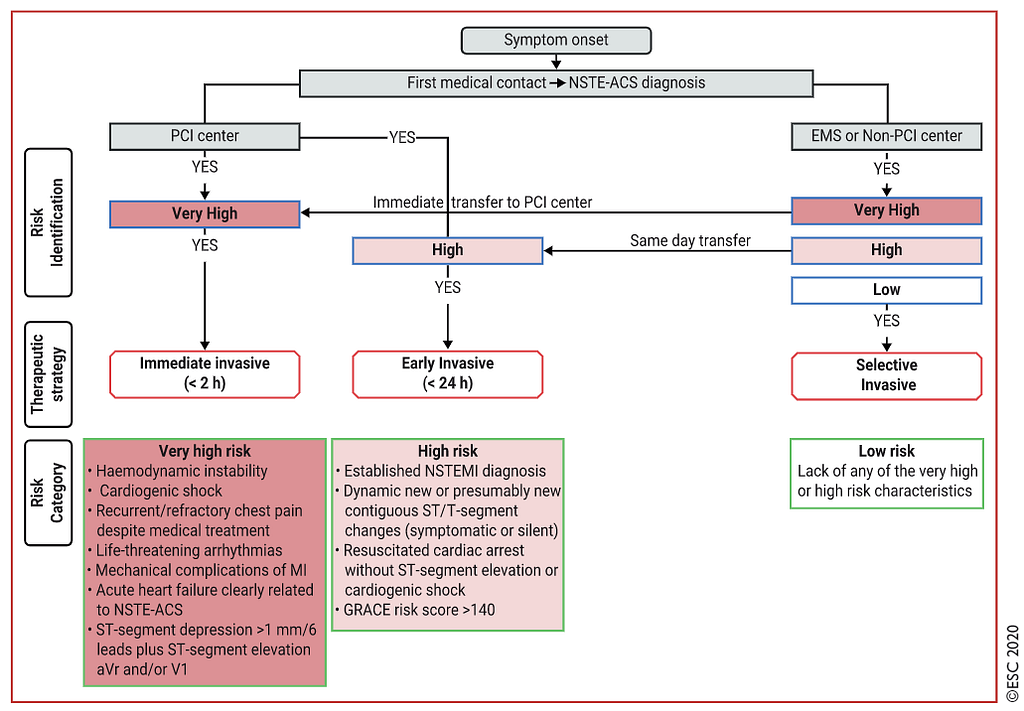
Remember the features of a very high risk P’t in the ESC 2020 NSTEMI guideline, the one we've talked to death? (see the bottom left of Fig.15)
Anyone who meets a Very high risk feature needs an immediate CV man consult. As for whether they "can" or "need to" go straight to the cath lab, that's not something we control. What the ED can control is giving the CV man as much of the information they need as possible: whether the patient is stable right now, whether there might be cardiogenic shock, whether they've gone into VT/Vf in the ED, whether there's Acute pul.edema, whether there are post-AMI mechanical complications. The more on-scene patient information we provide, the better the CV man can judge whether to activate the cath lab immediately.
After the consult, the CV man wanted to keep observing and wait for the biomarkers.

2.5 hr after arrival, the ECG showed RBBB+LAFB, with STE at the J point in V2.
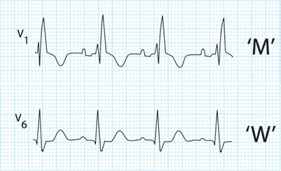
In RBBB morphology with a qR pattern or rsR’ pattern, where the right R’ > the left r, you usually see STD with TWI or an isoelectric baseline (Fig.17). Amal Mattu has stressed many times in the ECG Weekly teaching that in RBBB, any STE in any lead means you have to consider OMI. This is the ST opposition rule for RBBB → in RBBB, the ST segment shouldn't go in the same direction as the last part of the QRS. For example, look at Fig.18: this patient had chest pain, and we see a PVC with an RBBB pattern, with STE in V2/V3, while the last part of this QRS also points up. That violates the ST opposition rule, so you have to consider that ischemia has produced primary STTC.
In RBBB with STD in V1~V3 (especially with rSR’), the normal STD is at most Up to 1 mm (again, this only applies when there's an R’-wave); only when the R’-wave is very large, e.g. RVH, can it be > 1 mm. If it's more than 1 mm, an ST vector force is pulling it down, causing excessive discordant STD; the most common cause is Post.OMI9.

So what does RBBB+LAFB showing up actually mean?
First, this pattern (RBBB+LAFB) at least lets you diagnose LAD occlusion (because the LAD supplies the anterolateral walls, the RBB, and the LAF)
Dr. Smith ECG’s Blog mentions RBBB+LAFB in this post10 (actually it comes up in a lot of posts). The Blog cites a paper11, which describes that some very severe LAD occlusions or LM occlusions present as RBBB+LAFB → these cases have the highest risk of Vf, cardiogenic shock, and the highest in-hospital mortality (AMI for new RBBB alone has 18.8%).
- Smith has stressed many times that this ECG pattern is the OMI with the highest mortality
That said, occlusion in the LAD is the more common situation; occlusion in the LM is very rare, mainly because patients with an LM occlusion rarely survive long enough to get an ECG and an angiogram.
Also, the STE in RBBB+LAFB is very often downsloping STE; this is almost routine rather than the exception (see the example in Fig.19).
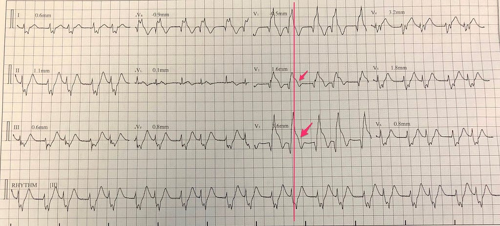
In Fig.19 the vertical line passes through the J point; V2/V3 both have obvious STE, and it's downsloping STE (see arrows).
With RBBB+LAFB it's not easy to tell there's a STEMI.
So Dr. Smith wrote a passage in this post12 → when others won't believe what we say, how do we persuade them? (I think there's a STEMI, but you think there isn't)
The original text:
『Stephen Smith:just use powers of persuasion. One is this: “the art of diplomacy is letting other people have your way” In other words, make them think it is their idea. Or make a plea for help: “I need your help on this.” People respond to being needed. If that fails, you say: “I am writing that I strongly believe this patient needs an emergent angiogram, right now. If you don’t do it, and you’re wrong, you will look very bad”』
My rough translation of the key meaning:
Let the other person (CV man) think it's their idea; also, say that I (ER Dr.) need your (CV man's) help. People usually want to help when they feel needed.
An example:
I think there might be a problem here on the ECG. Look, the J point is here, and the ST is already elevated. It seems like a STEMI. I need your help to take a look for me. (Turning our idea → into their idea is what gives it a chance to move forward)
This is a communication technique. Rather than flatly declaring this is a STEMI, point out the spots where you suspect OMI, let the CV man feel needed, and they'll be more willing to focus on the places you think may be a problem.
When I teach medical students ECG, I often say ECG reading, like POCUS, is operator dependent. An ECG comes up, and some people can see the ischemic change, while others just can't see it no matter what. It comes down to how much you practice. Only by constantly reading the masters' ECG blogs and practicing reading ECGs can you improve your accuracy with OMI ECG findings.
Back to the case again~~~
The patient had newly onset RBBB and then recurrent chest pain. The CV man still didn't want to do an emergent cath. Only after 3.5 hours, when the ECG went from RBBB back to something like the 1-hour ECG + ongoing symptoms + rising TnI (second time), did he go to the cath lab.

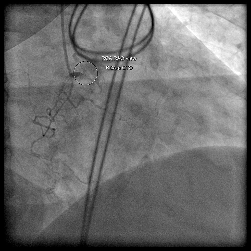
Cath report: LAD-p total occlusion, RCA-p CTO
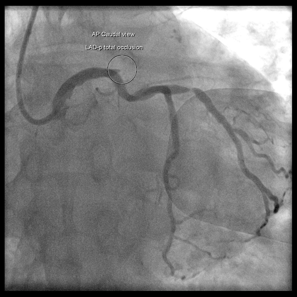
One last thing: which ECG findings support acute proximal LAD occlusion? (See this post13)
- STE starts in V1 ➡ with a mid or distal LAD occlusion, precordial STE typically starts in V2 or V3
- Marked STD reciprocal change in any of the inf.leads ➡ a mid or distal LAD occlusion is unlikely to show reciprocal STD change in the inf.leads
- STE in aVL ➡ this is unlikely in a mid or distal LAD occlusion (STE in Lead I can also mean Proximal LAD, but STE in aVL is the more reliable indicator for suspecting proximal LAD occlusion)
- When there's a new, evolving Ant.wall STEMI with new RBBB+LAFB or ICRBBB+LAFB, it's usually related to acute Proximal LAD occlusion (just like this case)
Also remember: RBBB+LAFB doesn't automatically mean LAD occlusion. It's clinical symptoms + newly onset of this ECG pattern that means you must strongly consider it.
Key points:
- Before using the Smith 4 variable formula, which ECG morphologies have to be excluded?
- In what situation does T/QRS ratio > 0.36 lean more toward STEMI?
- What do we do when the initial ECG is a non-diagnostic ECG? (What do ACLS 2020 and the 2022 ED acute chest pain consensus each say?)
- What are the Very high risk features in the ESC 2020 NSTEMI guideline?
- In RBBB, any STE or excessive discordance in V1–3 may mean OMI
- Key points on RBBB+LAFB → may be a proximal LAD occlusion; this pattern is the OMI with the highest mortality
- Which ECG findings support acute proximal LAD occlusion?
Additional references: 1 2 3 4 5 6 7 8 10 11 12 9 13
Thygesen, K., Alpert, J. S., Jaffe, A. S., Chaitman, B. R., Bax, J. J., Morrow, D. A., & White, H. D. (2018). Fourth Universal Definition of Myocardial Infarction (2018). Journal of the American College of Cardiology, 72(18), 2231–2264. https://doi.org/10.1016/j.jacc.2018.08.1038 ↩︎ ↩︎
Chest Pain, “Negative” Stress Tests, POCUS, & ECG Equations — REBEL EM — Emergency Medicine Blog — link ↩︎ ↩︎
O’Gara, P. T., Kushner, F. G., Ascheim, D. D., Casey, D. E., Chung, M. K., de Lemos, J. A., Ettinger, S. M., Fang, J. C., Fesmire, F. M., Franklin, B. A., Granger, C. B., Krumholz, H. M., Linderbaum, J. A., Morrow, D. A., Newby, L. K., Ornato, J. P., Ou, N., Radford, M. J., Tamis-Holland, J. E., … Zhao, D. X. (2013). 2013 ACCF/AHA Guideline for the Management of ST-Elevation Myocardial Infarction. Journal of the American College of Cardiology, 61(4), e78–e140. https://doi.org/10.1016/j.jacc.2012.11.019 ↩︎ ↩︎
Khawaja, M., Thakker, J., Kherallah, R., Ye, Y., Smith, S. W., & Birnbaum, Y. (2021). Diagnosis of Occlusion Myocardial Infarction in Patients with Left Bundle Branch Block and Paced Rhythms. Current Cardiology Reports, 23(12), 187. https://doi.org/10.1007/s11886-021-01613-0 ↩︎ ↩︎
Larson, D. M., Menssen, K. M., Sharkey, S. W., Duval, S., Schwartz, R. S., Harris, J., Meland, J. T., Unger, B. T., & Henry, T. D. (2007). “False-positive” cardiac catheterization laboratory activation among patients with suspected ST-segment elevation myocardial infarction. JAMA, 298(23), 2754–2760. https://doi.org/10.1001/jama.298.23.2754 ↩︎ ↩︎
Chang, A. M., Shofer, F. S., Tabas, J. A., Magid, D. J., McCusker, C. M., & Hollander, J. E. (2009). Lack of association between left bundle-branch block and acute myocardial infarction in symptomatic ED patients. The American Journal of Emergency Medicine, 27(8), 916–921. https://doi.org/10.1016/j.ajem.2008.07.007 ↩︎ ↩︎
Jain, S., Ting, H. T., Bell, M., Bjerke, C. M., Lennon, R. J., Gersh, B. J., Rihal, C. S., & Prasad, A. (2011). Utility of left bundle branch block as a diagnostic criterion for acute myocardial infarction. __The American Journal of Cardiology, 107(8), 1111–1116. https://doi.org/10.1016/j.amjcard.2010.12.007 ↩︎ ↩︎
Kontos, M. C., de Lemos, J. A., Deitelzweig, S. B., Diercks, D. B., Gore, M. O., Hess, E. P., McCarthy, C. P., McCord, J. K., Musey, P. I., Villines, T. C., & Wright, L. J. (2022). 2022 ACC Expert Consensus Decision Pathway on the Evaluation and Disposition of Acute Chest Pain in the Emergency Department. Journal of the American College of Cardiology, S0735109722066189. https://doi.org/10.1016/j.jacc.2022.08.750 ↩︎ ↩︎ ↩︎ ↩︎ ↩︎
Dr. Smith’s ECG Blog: Right Bundle Branch Block and ST Depression in V1-V3. Is that normal? And a complication. — link ↩︎ ↩︎
Dr. Smith’s ECG Blog: Cardiac Arrest at the airport, with an easy but important ECG for everyone to recognize — link ↩︎ ↩︎
Widimsky, P., Rohac, F., Stasek, J., Kala, P., Rokyta, R., Kuzmanov, B., Jakl, M., Poloczek, M., Kanovsky, J., Bernat, I., Hlinomaz, O., Belohlavek, J., Kral, A., Mrazek, V., Grigorov, V., Djambazov, S., Petr, R., Knot, J., Bilkova, D., … Lorencova, A. (2012). Primary angioplasty in acute myocardial infarction with right bundle branch block: should new onset right bundle branch block be added to future guidelines as an indication for reperfusion therapy? European Heart Journal, 33(1), 86–95. https://doi.org/10.1093/eurheartj/ehr291 ↩︎ ↩︎
Dr. Smith’s ECG Blog: What is the Diagnosis in this 70-something with Chest Pain? — link ↩︎ ↩︎
Dr. Smith’s ECG Blog: CO poisoning. Routine ECG recorded before hyperbaric therapy….Are they related? — link ↩︎ ↩︎



Thoughts on this case? Leave a comment, a like, or a reaction below.
comments powered by Disqus