Translated from the original Traditional Chinese post. Read the Chinese original →
Abdominal pain and chest tightness after drinking~~
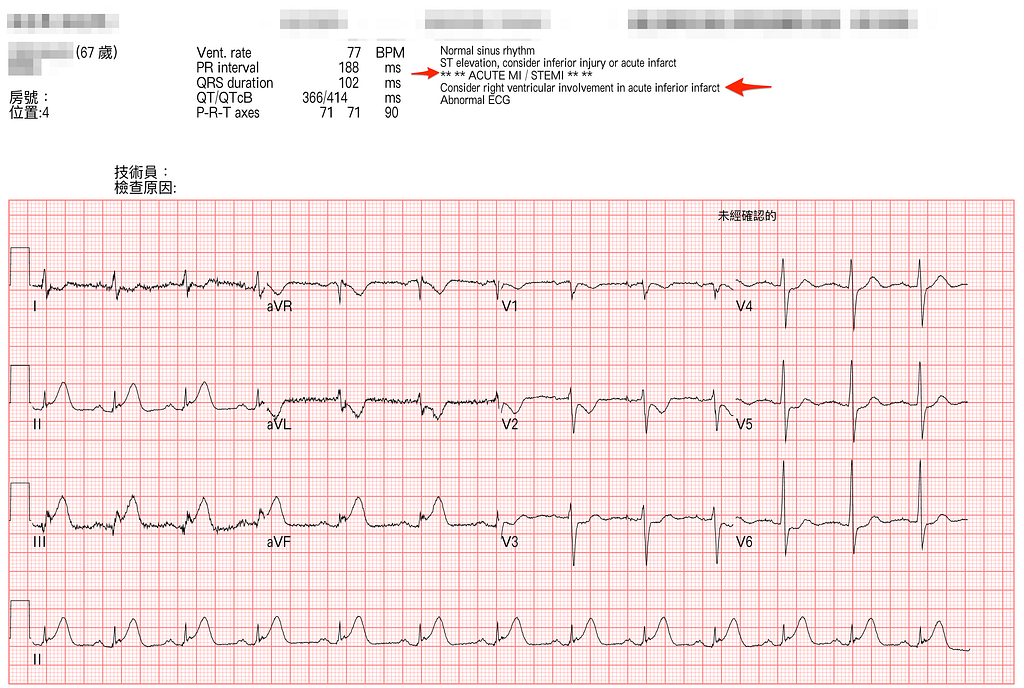
A 67-year-old man had a little kaoliang (Taiwanese sorghum liquor) at lunch, then started feeling chest tightness and epigastric pain~~~
As soon as the nurse finished the ECG, they told me right away: Dr. Bear~~~~ it's a STEMI~~~
ECG reading:
Rate:77 bpm, sinus rhythm, Normal axis, 1st degree AVB
Ischemia ECG findings are as follows:Hyperacute T wave in inf.leads with reciprocal STD change over aVL/I, STD in V1~V6(Maximal STD at V2~V3)
I want to point out a downsloping STD here (in this case → present in V1~V2, especially V2)
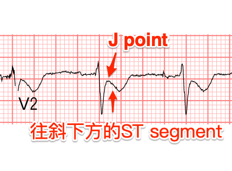
Dr.Smith stresses on his blog that any maximal STD in V1~V4 has to be considered post. OMI (96% specificity for Post.OMI) until proven otherwise. Especially downsloping STD in V2~V3: Smith says that in his experience, that alone is enough to diagnose post. OMI1.
Side topic: STD comes in three shapes (Fig.2)

Pay special attention: although upsloping STD leans non-ischemic, watch out for De Winter’s T wave → it has upsloping STD + hyperacute T wave, and the upsloping STD here is an STD that leans toward ischemic change.

So this case has an inf.-post. wall MI, with no obvious lateral wall involvement.
Here are a few fun questions:
Q1: When should you consider RVMI? How do you diagnose it? What's the blood supply?
Q2: Once you've done a right side ECG, how do you read it?
Q3: Why does identifying RVMI matter? (Where's the culprit lesion? Clinical management?)
Q4: In RVMI, is giving NTG really a no-go?
Let's go through these questions one at a time~~
Q1: When should you consider RVMI? How do you diagnose it?
When there's an accompanying inf. wall MI, consider whether there's also an RVMI.
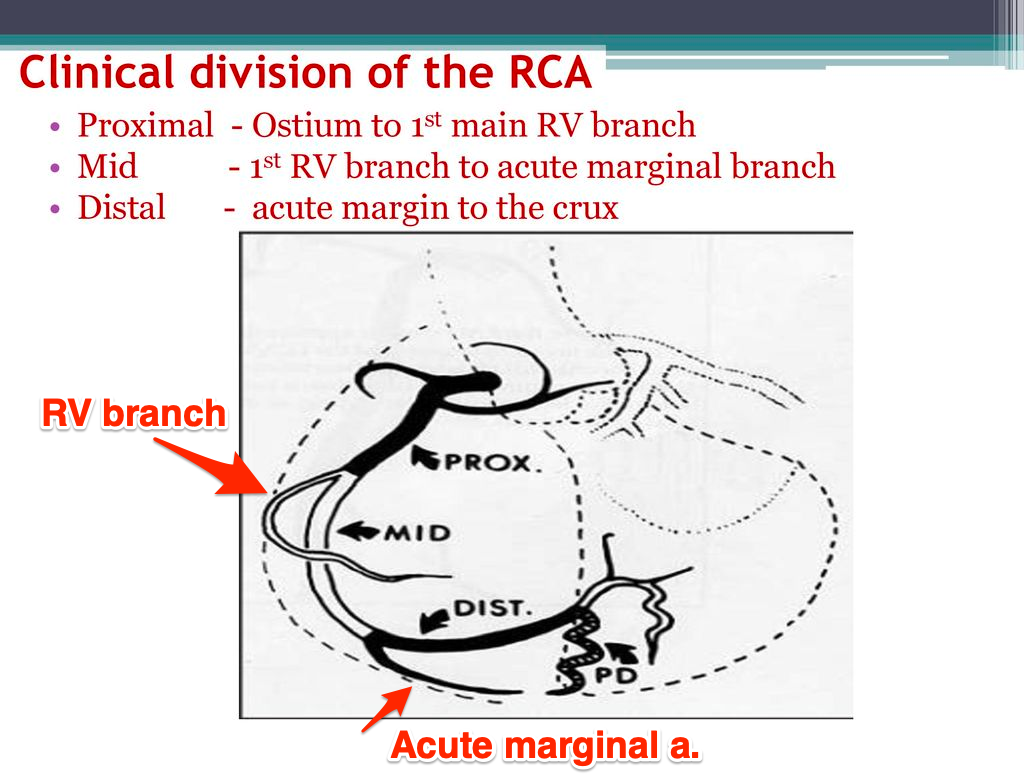
The RV's blood supply (Fig.3) comes from the RV branch of the RCA (also called the RV marginal branches). When we say most RVMI involves proximal RCA occlusion, we mean occlusion of the vessel before the RV branch. In other words, if you see RVMI, you infer that the RV supplied by the RV branch is affected, meaning the proximal RCA has a problem and blood can't reach the RV.
How can the ECG help with the diagnosis?
Either the regular 12-lead ECG or an R’t side ECG can help.
The easier one to understand is doing an R’t side ECG. That helps with the diagnosis.
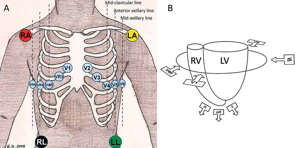
On the standard 12-lead ECG, V1 is the lead closest to the RV (Fig.4B), and it's the one better able to reflect ischemic changes in the RV. But overall, the standard 12-lead ECG isn't sensitive enough for diagnosing RVMI.
That also means we can't rule out RVMI with a 12-lead ECG → because V1 is the lead closest to the RV, when there's V1 STE (but no STE in the other ant. leads), together with chest pain with inf. wall STEMI → in this clinical scenario, strongly suspect RVMI
If you're using the 12-lead ECG to help decide, also strongly suspect RVMI if any of the following are present: (the ones marked ⭐️ are the more important ones)
- STD in V2, plus any one of the following (Amal Mattu especially emphasizes this)(⭐️)
➡︎STE in V1 →Highly specific for RVMI
➡︎ST isoelectric in V1
- STE in V1(⭐️)
➡︎If there's no STD in V2, STE in V1 has decent sensitivity and specificity for RVMI
➡︎If there is STD in V2, the sensitivity of STE in V1 drops (because it's harder to see → mainly because a posterior OMI pulls the RVMI's V1 STE down, so false-negatives come up easily)
- STE III >>> II (the much-greater-than part is what Amal Mattu emphasizes)(⭐️)
- STE in V1 > V2 (you could also call it a “Flattened” V2)(because in RVMI V1 rises, higher than V2)(⭐️)
- STE in aVR with inf. wall MI → should also hint at possible RVMI, so doing a right side ECG is a good idea, especially with every inf. wall MI
- STD and TWI in aVL →> 1 mm STD in aVL has 87% sensitivity and 90% PPV for acute inferior OMI with RVMI2
- STE in lead III>II, and STE in aVF greater than the degree of STD in V2
- STD in V2 ≤ 50% of the magnitude of STE in aVF34
- RBBB, 2nd or 3rd degree AV block
Those are ways to estimate, at first glance, whether there's RVMI when you see an inf. wall STEMI on a standard 12-lead ECG. But if the inf. wall STEMI isn't obvious and you still suspect RVMI, do an R’t side ECG; it usually gives more valuable information. Next, answering Q2~~
Q2: Once you've done a right side ECG, how do you read it?
Take a look at Fig.4A. The way we more commonly do an R’t side ECG is to keep V1/V2 in place and move V3~V6 to the mirror positions on the right chest wall. (as in Fig.4A)
If any one lead in V3R~V6R has STE > 0.5 mm, that counts as RVMI.
If there's no time for a full right side ECG → just do V4R (V4R is considered the best lead for diagnosing RVMI)5
STE ≧ 1 mm in V4R has 88% sensitivity and 78% specificity for RVMI6
Also, reading a right side ECG is like V2~V3 in the STEMI criteria: you need to pay attention to sex/age. Here it is all in one table: (Fig.5 → STEMI definition, post. leads definition, R’t side ECG definition)
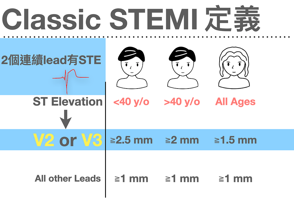
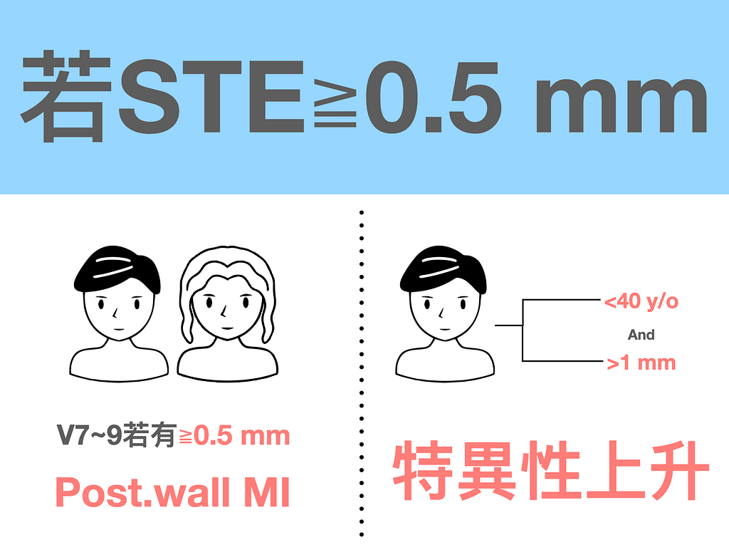
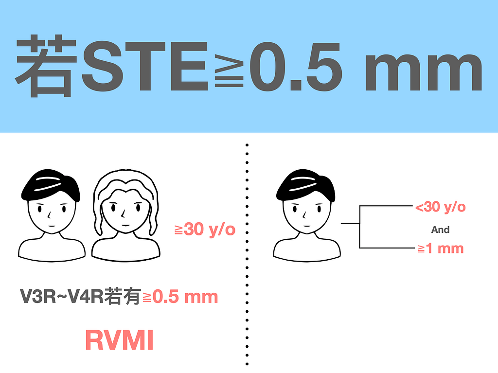
Don't overthink it. Just remember that for these extra leads (post. leads/R’t side ECG), the STE cutoff is 0.5 mm, and in younger patients the STE cutoff is 1 mm.
There's one more important point about the R’t side ECG: do it early.
Why? Because R’t side ECG STE from RVMI is transient. This study found that in 50% of acute RVMI patients, the right side lead STE resolves within 12 hours of ACS S/S onset7.
So what affects STE in V3R and V4R?8
- If the STE in the inf.leads isn't obvious, expect it not to be very obvious on the right side ECG either
- V3R and V4R look at rightward, inward vectors; if the case also has an MI with lateral wall extension, they cancel each other out, making the STE in V3R/V4R less obvious
- STE suggests transmural ischemia rather than infarction
Q3: Why does identifying RVMI matter? (Where's the culprit lesion? Clinical management?)
About 25% of inf. wall MIs have an accompanying RVMI. From Fig.3 we know that the RV's blood supply comes from the RV branch of the proximal RCA (the LCx doesn't supply the RV). So when we see RVMI, we guess the culprit lesion is in the proximal RCA.
Diagnosing RVMI affects clinical management, because the clinical course, prognosis, and hemodynamic treatment of acute RVMI are clearly different from LVMI (e.g., hypotension, relative hypovolemia, hypersensitivity to NTG)9
These patients with RVMI are very sensitive to preload (because RV contractility is poor), so nitrates or other preload-reducing drugs may drop their blood pressure too low.
Recognizing RVMI also matters because it comes with higher short-term morbidity and mortality10
- Inf.wall STEMI with/without RVMI involved ➡ with RVMI, both serious complications (45% vs. 19%) and death rates (22% vs. 6%) are higher
There's also a concept here you may need to know.
In general, most proximal RCA occlusions don't readily show STE in V1, mainly because in many people the RV's blood supply comes not only from the RV branch but also from collateral circulation coming over from the LAD. So even if flow in the RV branch is cut off, there's still supply from the LAD.
1️⃣If you see STE in V1 or R’t side ECG STE ➡ favor RVMI, and you can infer proximal RCA occlusion
2️⃣If there's no ECG evidence for RVMI ➡ you can't say there's no proximal RCA occlusion (because there may be collateral circulation from the LAD supplying it, so it doesn't lead to RVMI)
Q4: In RVMI, is giving NTG really a no-go?
First, to stress it again: RVMI is mainly caused by proximal RCA occlusion proximal to the RV branch, leading to RV ischemia. This happens especially when there's no collateral circulation from the LAD supplying the RV.
So why do we worry about RVMI ➡ because it can lead to RV failure, shock, hypotension
But…. and it's exactly this but……
Only a minority of RVMIs have this kind of hemodynamic instability, and even fewer proximal RCA lesions would do this, because most have collateral circulation from the LAD protecting the RV. So seeing RVMI doesn't automatically mean the RV will dilate and fail, with NTG making things worse. Unless the collateral circulation isn't pulling its weight11.
So it's when there's hemodynamic instability that diagnosing RVMI becomes more important.
The case continues
After all that, let's look at the computer read on Fig.1 → Acute STEMI, RV involved in inf.MI
Spot on~~~~
Let's apply the truckload I wrote above about using a regular 12-lead ECG to work out whether there's RVMI to this ECG.
With STD in V2, is there STE in V1? → No, but it looks close to the isoelectric baseline
Also STE in III >>>II, and there's also STE in aVR
This patient got an R’t side ECG right away (Fig.6).

The R’t side ECG showed no STE in V3R or V4R, but V6R had STE (>1 mm)
So this case has an inf.-post. wall MI with an accompanying RVMI.
CAG:Culprit lesion at RCA-m stenosis
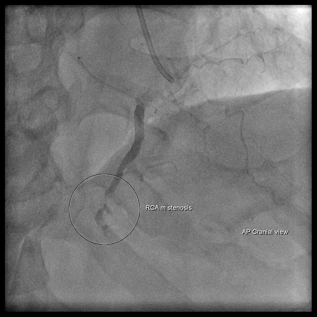
The lesion was at RCA-m~~~~~~ but wasn't there an RVMI? Why?
I don't know the reason either.
In a YouTube conversation between Smith and Tom Bouthillet, I saw a case with a similar situation.

At 7:10 in the video, Smith explains that it's also possible the thrombus was originally in the proximal RCA, but when the PCI went in, the thrombus turned into emboli and lodged in RCA-m.
Keypoints:
- When downsloping STD shows up, watch out for ischemia!!!
- The three shapes of STD
- How to diagnose RVMI with a regular 12-lead ECG, and how to diagnose it with an R’t side ECG
- Why RVMI matters
Additional references: 1 2 3 5 6 7 9 8 11 10
Dr. Smith’s ECG Blog: Any ST depression in V2 and V3 is posterior OMI until proven otherwise, especially if downsloping — link ↩︎ ↩︎
Turhan, H., Yilmaz, M. B., Yetkin, E., Atak, R., Biyikoglu, S. F., Senen, K., Ileri, M., Cehreli, S., Korkmaz, S., & Kutuk, E. (2003). Diagnostic Value of aVL Derivation for Right Ventricular Involvement in Patients with Acute Inferior Myocardial Infarction. __Annals of Noninvasive Electrocardiology__, __8__(3), 185–188. https://doi.org/10.1046/j.1542-474X.2003.08303.x ↩︎ ↩︎
Somers, M. P., Brady, W. J., Bateman, D. C., Mattu, A., & Perron, A. D. (2003). Additional electrocardiographic leads in the ED chest pain patient: right ventricular and posterior leads. __The American Journal of Emergency Medicine__, __21__(7), 563–573. https://doi.org/10.1016/j.ajem.2003.08.008 ↩︎ ↩︎
Lew, A. S., Laramee, P., Shah, P. K., Maddahi, J., Peter, T., & Ganz, W. (1986). Ratio of ST-segment depression in lead V2 to ST-segment elevation in lead aVF in evolving inferior acute myocardial infarction: an aid to the early recognition of right ventricular ischemia. The American Journal of Cardiology, 57(13), 1047–1051. https://doi.org/10.1016/0002-9149(86)90672-7 ↩︎
Carley, S. D. (2003). Beyond the 12 lead: Review of the use of additional leads for the early electrocardiographic diagnosis of acute myocardial infarction. __Emergency Medicine Australasia__, __15__(2), 143–154. https://doi.org/10/b344h9 ↩︎ ↩︎
Zehender, M., Kasper, W., Kauder, E., Schönthaler, M., Geibel, A., Olschewski, M., & Just, H. (1993). Right ventricular infarction as an independent predictor of prognosis after acute inferior myocardial infarction. __The New England Journal of Medicine__, __328__(14), 981–988. https://doi.org/10/bgmbpd ↩︎ ↩︎
Levis, J. (2016). ECG Diagnosis: Right Ventricular Myocardial Infarction. __The Permanente Journal__. https://doi.org/10.7812/TPP/16-105 ↩︎ ↩︎
Chhapra, D. A., Mahajan, S. K., & Thorat, S. T. (2013). A study of the clinical profile of right ventricular infarction in context to inferior wall myocardial infarction in a tertiary care centre. __Journal of Cardiovascular Disease Research__, __4__(3), 170–176. https://doi.org/10.1016/j.jcdr.2013.04.003 ↩︎ ↩︎
Dr. Smith’s ECG Blog: What is a useful next step in the evaluation of this patient with Chest pain and this ECG? — link ↩︎ ↩︎
Bischof, J. E., Worrall, C. I., & Smith, S. W. (2018). In inferior myocardial infarction, neither ST elevation in lead V1 nor ST depression in lead I are reliable findings for the diagnosis of right ventricular infarction. __Journal of Electrocardiology__, __51__(6), 977–980. https://doi.org/10.1016/j.jelectrocard.2018.08.010 ↩︎ ↩︎
Dr. Smith’s ECG Blog: Inferolateral STEMI: is there right ventricular MI also? — link ↩︎ ↩︎



Thoughts on this case? Leave a comment, a like, or a reaction below.
comments powered by Disqus