Translated from the original Traditional Chinese post. Read the Chinese original →
Can You See What's Wrong with This ECG?
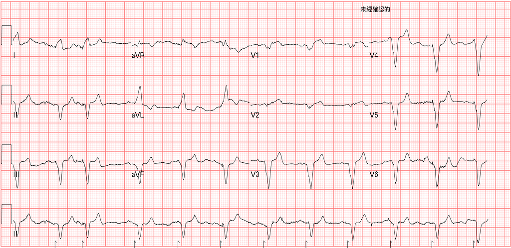
A 74-year-old man with chest pain, hurting so bad he almost passed out. On arrival he said it had been happening a lot over the past few days but always eased off quickly; today it was especially bad.
The ECG machine's interpretation usually isn't very trustworthy, but ECG machines are very sensitive at detecting a pacemaker rhythm1. Fig.1 is the ECG done within 10 minutes of arrival, and in the long lead II along the bottom row you can see the tiny pacemaker spikes it picked up.
So the question comes back to this: can you diagnose AMI on the ECG in a patient with a PPM?
So let's look at a paper that was already available in Annals of Emergency Medicine as an Article in press in June this year, and was formally published in October.

This paper uses the Modified Sgarbossa criteria to help diagnose ACO (Acute coronary occlusion) in patients with a pacemaker2.

In Fig.2 you can see the study had three groups (see Table 1 for the numbers)
❶The first group was OMI (59 patients)
❷The second group was the NOMI control group (90 patients)
❸The third group was the No OMI control group (102 patients)—pacemaker patients in the ED without OMI (who came to the ED with an ACS complaint)

OMI group: TIMI flow 0~1 or TIMI flow 2~3 + High peak Troponin
- 31 patients were diagnosed by Concordant STE or STD
- 2 patients were diagnosed by the > 5 mm rule of the Original Sgarbossa criteria
- 17 patients were diagnosed by the MSC C rule
So with MSC A+B+modified C➡you catch: 31 + 17=48➡48/59=81% (11 still missed) →this is the sensitivity of MSC for OMI
With Original SC A+B+C➡you catch: 31 + 2=33➡33/59=56% (26 still missed) →the sensitivity of Original SC for OMI
NOMI group (Control group)➡TIMI flow 2~3 + Low peak Troponin
Again, let's use MSC and Original SC and see what the false positives look like.
The false positive rate of MSC: in the NOMI group, 14 of the 90 patients were MSC(+) →14/90=16% (so Specificity is 84%)
The false positive rate of Original SC: 9 of the 90 patients were Original SC(+) →9/90=10% (so Specificity is 90%)
LR- →0.22 vs. 0.49 (MSC vs. OSC)
LR+ →5.2 vs. 5.6 (MSC vs. OSC)
No OMI group (Control group)➡PPM patients in the ED without OMI, collected at random
The false positive rate of MSC in the No OMI group: 4 of the 102 patients were MSC(+) →4/102=4% (so Specificity is 96%)
The false positive rate of Original SC: 3 of the 102 patients were Original SC(+) →3/102=3% (so Specificity is 97%)
LR- →0.19 vs. 0.45 (MSC vs. OSC)
LR+ →21 vs. 19 (MSC vs. OSC)
Putting the OMI group together, we find that MSC has better sensitivity for diagnosing OMI, as high as 81%. The Original SC's sensitivity for OMI is only 56%.
On top of that, the other two control groups (NOMI and No OMI) tell us that for patients without OMI, both MSC and Original SC have a very low false positive rate (that is, very high specificity (84%~97%) →Specificity=1-false positive rate). In other words, if MSC or Original SC is positive, you can rule in OMI.
Also, in the No OMI group there were 4 MSC(+) patients (4 of 102). When they analyzed these 4, they found these 4 patients' baseline ECGs were already MSC(+). This also teaches us that if an ACS patient with a pacemaker is MSC(+) in the ED, please also pull out their old ECGs and look at them together—it might be an old change.
Back to the case from the beginning
When I saw this arrival ECG in Fig.1 in the ED, I wrote on the ECG: V1 possibly MSC (meaning Criteria C →excessive discordant > 0.25)
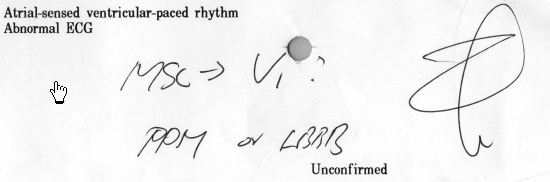
So I parked the patient right in front of my desk, on an ECG monitor. Because I thought the patient had a problem, because of MSC(+).
For MSC criteria A and C, any single lead meeting the rule counts as a problem. Criteria B: in LBBB you only look at V1~V3, but in a pacemaker patient you look at V1~V6.
40 minutes later, since the patient's symptoms persisted, we repeated the ECG (Fig.3).
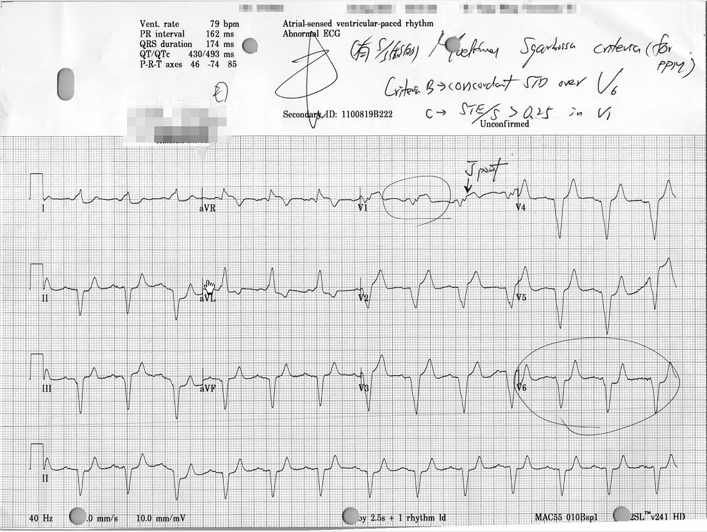
In Fig.3, V1 is still excessive discordant >0.25, and V6 shows concordant STD. Seeing that kind of change in V6
Another 20 minutes later, another ECG (still symptomatic) (Fig.4)
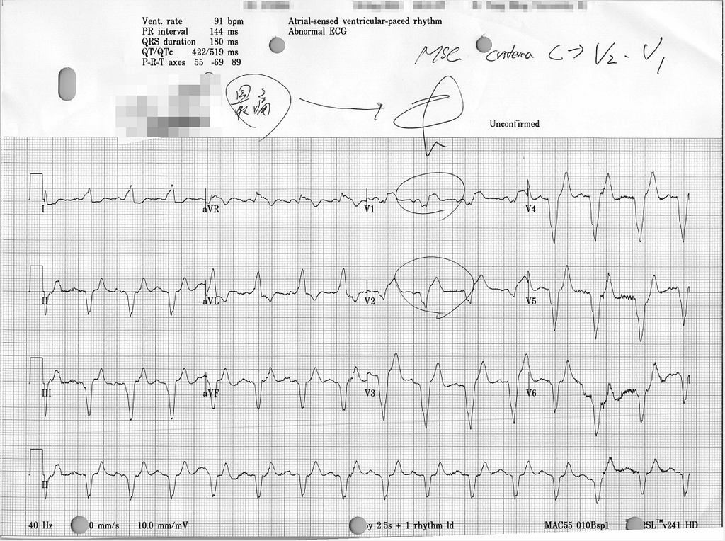
In Fig.4, both V1 and V2 meet MSC criteria C. In V5~V6 the baseline is unstable, so you can't tell whether there's concordant STD.
At this point, the CV man was finally willing to take the patient for PCI.
For the last few decades, MI patients have been split into STEMI/Non-STEMI. Everyone treats rushing a STEMI to the cath lab the moment you see it as the supreme guiding principle. But what often gets overlooked is: is ST elevation really that sensitive for MI?
Let's look at the cases collected in this paper3.

The paper collected 808 admitted ACS patients. 265 of them had an occluded vessel (meeting Occlusion MI), of whom 108 (41%) met STEMI criteria and 157 (59%) did not meet STEMI criteria.
So ST elevation actually has really poor sensitivity for diagnosing OMI. In other words, if you rely on ST elevation to catch OMI patients, you'll miss a lot of OMI.
In LBBB/PPM patients, the abnormal conduction alters ventricular depolarization and repolarization, which produces secondary STTC, and these STTC sometimes mask or mimic ischemic changes. That's why LBBB/PPM patients have long been considered hard to diagnose OMI in.
But now, for LBBB and pacemaker patients, validation studies have found that MSC can show the patient has OMI with very good sensitivity and specificity.
The case continued: PCI
CAG: LAD-M total occlusion (CTO) s/p stent, but failed, RCA-M ulcerative plaques with 80% stenosis s/p stent
Here let's look at a few questions.
❶First: can MSC localize the ischemic territory?
Normally, with normal conduction, we can infer the ischemic territory and the culprit lesion from the ECG. But even with the abnormal conduction of LBBB/PPM, we can still infer the likely location of ischemia from the leads that meet MSC criteria A, B, or C.
For example, Criteria A means concordant STE; in the leads with concordant STE, you may need to consider whether there's ischemia. In this case, V1~V2 met excessive discordant >0.25 (Criteria C), so you have to think about a septal wall problem. If there's Concordant STD over V1~V3 meeting Criteria B, you have to think about Post.wall OMI.
In other words, MSC can localize the infarcted/ischemic territory.
❷Second: since the patient showed ischemic changes in V1~V2, we'd guess it was an LAD problem. So why, when the LAD stent failed, did the leads for the LAD territory improve? (The RCA stent was successful—so how did the LAD ischemic leads go back to normal? →see Fig.5)
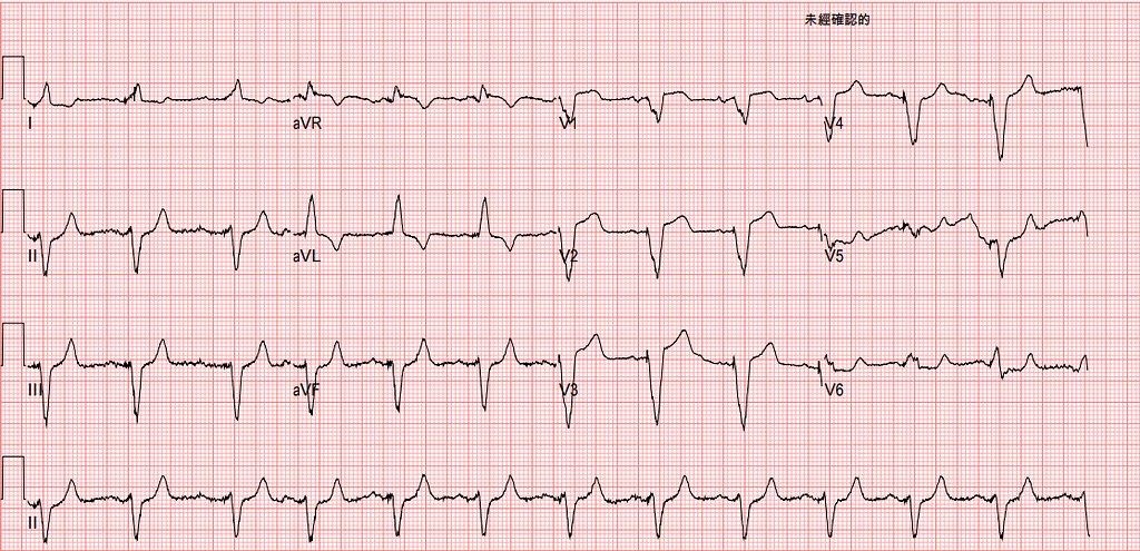
Fig.5 is the Post PCI ECG after the RCA stent (they tried to stent the LAD-CTO but couldn't open it). You can see that the excessive discordant STE (>0.25) in V1~V2 is gone. That means the ischemia improved.
But the LAD clearly couldn't be opened because of the CTO, and it was the RCA that got stented—so why did V1~V2, supplied by the LAD, improve?
I honestly couldn't figure this one out! So I went to twitter and asked some ECG masters~~~

After reading the masters' replies, it kind of clicked. How so?
The ECG actually can't truly localize the culprit lesion; it can only show the ischemic territory. Because the ischemic territory is confounded by the anatomy of the coronary arteries, whether there was a prior MI, whether there is collateral flow, and so on.
That is, take this example. The ECG looked like the septal wall might be in trouble. The root cause wasn't actually a problem in the LAD itself, but in the collateral circulation supplying the LAD-CTO territory (coming from the RCA). Before PCI, we'd have no way of knowing that at all. We'd only know the septal wall might be ischemic, but not who the real Culprit lesion was. Especially when the patient has disease in multiple vessels.
Only after PCI did we learn the LAD had a CTO, with collateral flow from the RCA helping supply it. When the RCA also has a problem, the RCA collaterals supplying the CTO territory are of course affected too. So once the RCA was fixed, the collaterals got better too, and naturally the OMI ECG findings in V1~V2 improved.
Learning points:
- For whether an LBBB/PPM patient has OMI, MSC can help with the diagnosis. Both Sensitivity and specificity are high
- The sensitivity of ST elevation for diagnosing OMI is less than half (so we should learn other OMI ECG findings all the more)
- MSC can localize the infarcted/ischemic territory, but cannot localize the culprit lesion
- The ECG can't definitively tell you which vessel the culprit lesion is in (most of the time it can). But knowing which one is the culprit lesion isn't important; what matters is knowing whether the patient has OMI. Because if there's OMI, they need PCI or another procedure to open the vessel.
Dodd, K. W., Zvosec, D. L., Hart, M. A., Glass, G., Bannister, L. E., Body, R. M., Boggust, B. A., Brady, W. J., Chang, A. M., Cullen, L., Gómez-Vicente, R., Huis in ‘t Veld, M. A., Karim, R. M., Meyers, H. P., Miranda, D. F., Mitchell, G. J., Reynard, C., Rice, C., Salverda, B. J., … Taylor, A. M. (2021). Electrocardiographic Diagnosis of Acute Coronary Occlusion Myocardial Infarction in Ventricular Paced Rhythm Using the Modified Sgarbossa Criteria. __Annals of Emergency Medicine__, S0196064421002493. https://doi.org/10.1016/j.annemergmed.2021.03.036 ↩︎ ↩︎
Meyers, H. P., Bracey, A., Lee, D., Lichtenheld, A., Li, W. J., Singer, D. D., Rollins, Z., Kane, J. A., Dodd, K. W., Meyers, K. E., Shroff, G. R., Singer, A. J., & Smith, S. W. (2021). Ischemic ST‐Segment Depression Maximal in V1–V4 (Versus V5–V6) of Any Amplitude Is Specific for Occlusion Myocardial Infarction (Versus Nonocclusive Ischemia). __Journal of the American Heart Association__, e022866. https://doi.org/10/gngnv7 ↩︎ ↩︎



Thoughts on this case? Leave a comment, a like, or a reaction below.
comments powered by Disqus