Translated from the original Traditional Chinese post. Read the Chinese original →
OHCA arrives at the ED: do we activate the cath lab?

On one dark and stormy night, the fire department EMS radio outside the ED crackled:
This is Yilan 91, we're bringing in a medical OHCA to NYCU Hospital (National Yang Ming Chiao Tung University Hospital) shortly.
The ambulance arrived: a middle-aged woman, roughly 50 years old, pale, with no color in her face at all.
The 91 crew told us the AED on scene advised no shock, and they had kept doing CPR.
In the resus room we put her on the ECG monitor. No palpable pulse, but there was an ECG waveform; it looked like PEA. After a few rounds of compressions and drugs, the nurse called out that we'd gained a pulse.
We immediately got the 12-lead ECG in Fig.1.
Let's read this ECG:
Rate:150 bpm
Rhythm:Upright P wave in II>I, inverted P wave in aVR, so this should be sinus rhythm
Axis:Normal axis
Interval:QTc →507 ms
Ischemia:We see multiple-lead STD + aVR and/or V1 STE (Fig.2)

A few questions worth thinking through:
Q1: What kind of situation causes ECG changes like this?
Q2: What conditions give you rSr’ in V1-V2?
Q3: Is there a problem with this QTc value?
Q4: What are the key points to watch after ROSC in an OHCA patient?
Q5: If we suspect ACS, do we rush to the cath lab? What do we need to rule out before going?
First, when you see diffuse STD + aVR and/or V1 STE, the most common problem is an oxygen demand and supply mismatch causing subendocardial ischemia. One feature of this kind of STD is that it can't localize the ischemic territory. Take Fig.2 as an example: we see STD in II/III/aVF and V3~V6, but we can't say the patient has ischemia of the inf. wall or ant./lateral wall, because subendocardial ischemia usually can't localize where the ischemia is.
Flip it around: what kind of STD can localize the ischemia? When the STD appears in the territory reciprocal to a vessel's distribution, rather than obvious diffuse STD, it can reflect where the heart is ischemic. And what it reflects is ischemia in the leads opposite to where the STD shows up (the STD here means reciprocal change → this is very, very important).
👉V1~V4 and V7~V9 are reciprocal to each other:
- If we see maximal STD in V1~V4, the first thing to think of is post. wall ischemia → V1~V4 and V7~V9 face each other
👉I/aVL and II/III/aVF are reciprocal to each other:
- If we see STD in I/aVL, think of the opposite leads (inf. wall ischemia)
- If we see STD in II/III/aVF, think of the opposite leads (high lateral wall ischemia)
👉V1/V6 are reciprocal to each other:
- If we see STE in V1 with STD in V5–6, think of septal wall ischemia
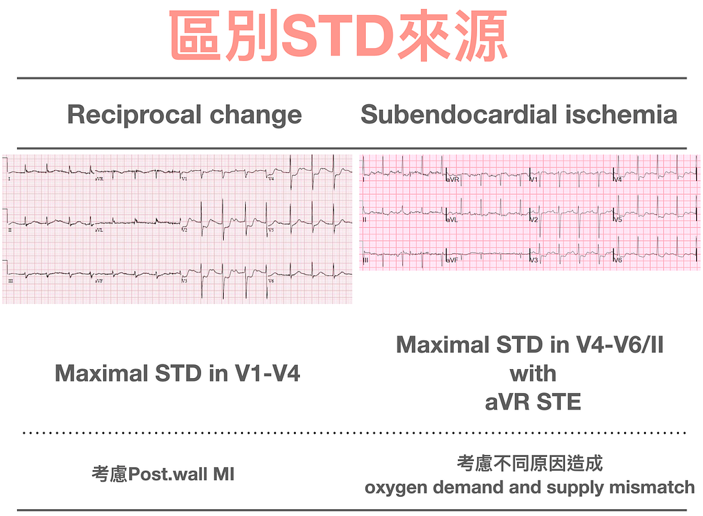
ECG Tips:How do we tell whether STD is subendocardial ischemia or reciprocal change? See Fig.3. If the maximal STD is in V4–6/II with aVR STE, think subendocardial ischemia first; if the STD is confined to a vascular territory, consider reciprocal change first
But there are way too many causes of oxygen demand and supply mismatch that produce the subendocardial ischemia ECG pattern; see Table 1. I've posted this table many times. Besides ACS sick P’t, where it could be LMCA, TVD, or proximal LAD, there are even more non-ACS sick P’t who also show this ECG pattern. So when we see the subendocardial ischemia ECG pattern, the only trick is a good physical exam to look for more clues, not blindly deciding the patient is an ACS sick P’t.
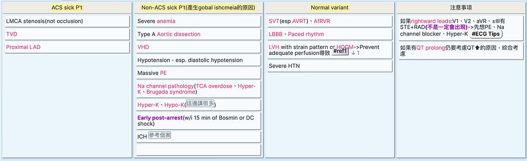
ECG Tips:Don't treat a subendocardial ischemia ECG pattern like you just found a loaded gun and fire it straight at ACS, because many more causes can produce this pattern
Next, the second question: what situations produce an rSr’ pattern in V1-V2?
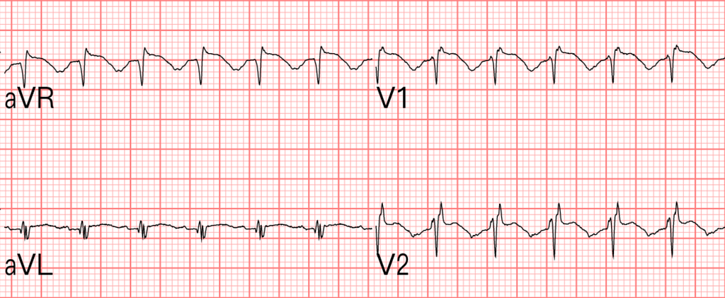
In general, the P wave in V1 is mostly upright or biphasic (with the inverted part not prominent). When you see an obviously inverted P wave, consider that V1–2 were placed too high.
In this post, the master Ken Grauer notes that if you see the following clues, you should consider that V1/V2 were placed too high1.
👉A septal Q wave may show up because V1/V2 were placed too high
- Although a deep Q wave or QS wave in V1/V2 should make you consider septal infarction, if you don't also see a Q wave or QS wave in V3 ➡ most of the time it's not septal infarction (meaning that if there really is a septal infarction, you'd see Q waves or QS waves in V1~V3 together)
👉If V1 and/or V2 show an r’ wave, especially with ICRBBB ➡ it's more commonly due to the leads being placed too high
👉If V1 and/or V2 show an obvious negative component of the P wave
- When V2 is placed too high, atrial depolarization heading toward the apex moves away from V2 (because V2 is placed high)
- The P wave in V2 should be upright; if you see it inverted ➡ V2 is very likely placed too high
- A normal negative component of the P wave in V1/V2 is usually not very prominent
👉If V1/V2 look a lot like aVR, also consider that they were placed too high
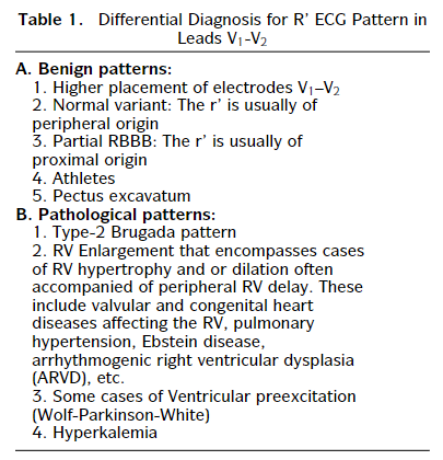
This article also discusses which DDx to consider further when V1-V2 show rSr’ (Fig.5)2.
Reading the original ECG (Fig.4), we see ICRBBB, rSr’ over V1, and V1 looking a lot like aVR, so we judged this to be V1/V2 misplacement, from the leads being placed too high.
The next question: at an H.R of 150, judging the QT interval is genuinely not easy.
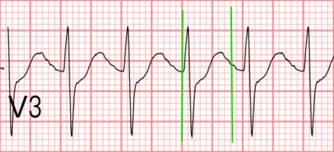
If you want to measure the QT interval, pick the longest one among the 12 leads, usually V2~V3 (Fig.6)
Plugging the roughly 320 ms QT interval from Fig.6 into the formula, we get a QTc of 508 ms
Corrected QT Interval (QTc) - MDCalc
Honestly, the whole reason we look at the QT interval is the fear that QT prolongation will tip over into TdP. If you don't want to calculate the QTc
you can use the QT nomogram (Fig.7)
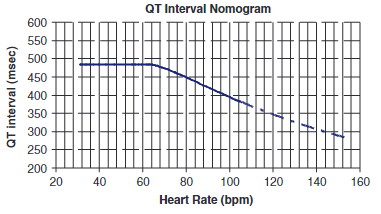
What Fig.7 shows is that a QT interval above the blue line carries a possibility of going into TdP:
- H.R<60 → use 485 ms (12 small boxes)
- H.R>100 → use 400 ms (10 small boxes)
If you keep those two numbers in mind, you can quickly assess whether there's a risk of TdP without calculating the QTc.
Of course, nowadays nearly every 12-lead ECG machine calculates the QTc for you. When you see QTc>500 ms, be careful.
With QTc>500 ms, watch for two broad groups of pathophysiology causing QT prolongation:
- QT prolongation from a bigger T wave and QT prolongation from a longer ST segment (see Fig.8)

ECG Tips:QTc>500 ms is a problem; H.R<60 → QT interval>12 small boxes, and H.R>100 → QT interval>10 small boxes, may both be a problem
Next, let's look at the key points to watch after ROSC in an OHCA patient.
First, what does post-cardiac arrest care look like in ACLS 2020? (Fig.9)
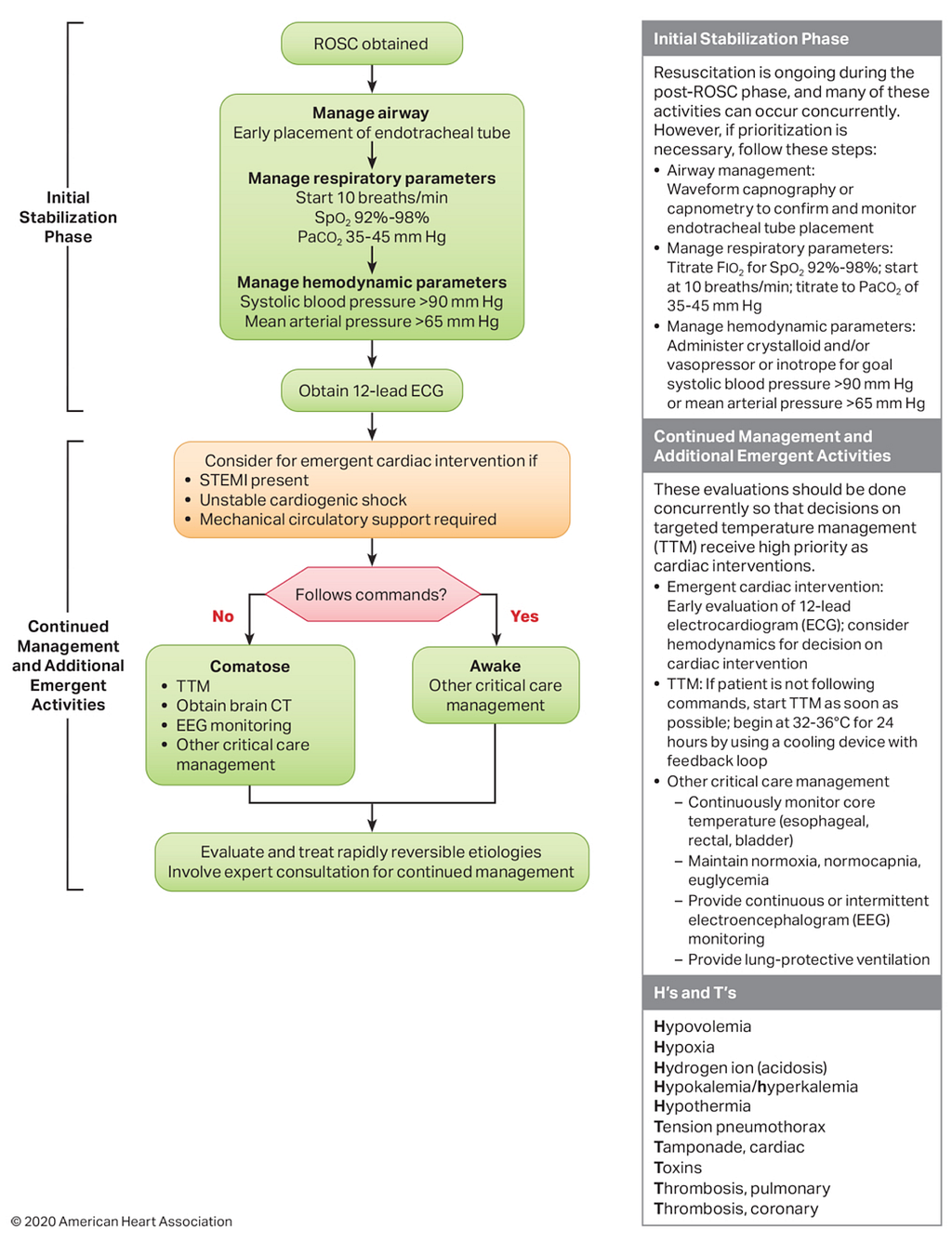
Management after ROSC comes in two parts:
👉Initial stabilization phase
1.Manage airway (A)
- Early placement of ET tube→use waveform capnography or capnometry to confirm ET tube position
2.Manage respiratory parameters (B)
- Start 10 breaths/min
- Titrate FiO2→Keep SaO2→92~98%
- PaCO2 35~45 mmHg
3.Manage hemodynamic parameters (C)→give crystalloid ±vasopressor
- SBP>90 mmHg and MAP>65 mmHg
Get a 12-lead ECG after A, B, and C are stabilized
👉Continued management and additional emergent activities (PCI/TTM/other treatment)
1.Consider PCI if any of the following
- STEMI(+)
- Unstable cardiogenic shock
- Patients who need mechanical circulatory support (meaning IABP, ECMO, etc.)
2.Can the patient follow commands? (to decide whether to use TTM)
- Awake→other critical care management
- Comatose→TTM, get a brain CT, EEG monitoring
TTM:
- Begin at 32~36°C(Class I ) for 24 hrs(Class IIa)
- Whether IHCA or OHCA, and whatever the initial rhythm, TTM is recommended for anyone with ROSC (Class I)
- Routine use of cold IV fluids for patients with prehospital ROSC is not recommended (Class No benefit)
3.Critical care management
- Continuously monitor core temperature(esophageal, rectal, bladder)
- Maintain normoxia, normocapnia, euglycemia
- Provide continuous or intermittent EEG monitoring
- Provide lung-protective ventilation
Tips:After ROSC, correct abnormalities step by step following Airway, Breathing, Circulation, then get a 12-lead ECG to decide whether to rush to the cath lab, and then proceed with additional emergent management based on the patient's level of consciousness
So, is this case a good candidate for the cath lab?
Let's take a closer look at what ACLS 2020 actually says about whether ROSC patients should get a cath.
In OHCA with ROSC, the role of CAG is still under discussion. When should we activate the cath lab? This 2019 Circulation article describes the following3:
- Patients whose OHCA was due to a shockable rhythm (Vf or pulseless VT) have a high chance that severe CAD caused the cardiac arrest ➡ in these patients, the prevalence of CAD is 70~85%
- If the post-ROSC ECG shows STE, the prevalence of CAD can be as high as 96% ➡ immediate cath lab activation gives better survival to hospital discharge
- In patients without STE after ROSC, the prevalence of CAD is still 25~50%, and in these patients cath lab activation still offers some benefit

Fig.10 describes when PCI is recommended in cardiac arrest:
- Patients with suspected cardiac-cause collapse, and patients whose post-ROSC ECG shows STE: both groups should get immediate CAG ➡ Class I
- Patients with suspected cardiac-cause OHCA with ROSC who are comatose and have no STE on the ECG, in some patients such as those with hemodynamic instability or who go into VT/Vf (electrical instability) ➡ it's also reasonable for these patients to get CAG ➡ Class IIa
- When a ROSC patient meets the criteria for CAG ➡ regardless of mental status ➡ Class IIa
In addition, if the patient is comatose after OHCA with ROSC, there's evidence that CAG is as beneficial as it is in patients who are awake ➡ so whether to do CAG has nothing to do with the patient's neurologic status (in other words, whether to do CAG has nothing at all to do with whether the patient is awake or not; if they meet the indication, you can go).

This figure shows the key points in the 2015 ACLS and 2020 ACLS post-cardiac arrest algorithms about whether to do a cath.
- If there's STEMI after ROSC, the guideline says rush to the cath lab
- Suspected cardiac-cause collapse also means rush to the cath lab (for example, the patient complained of chest pain before going down)
ECG Tips:STE after ROSC, suspected cardiac-cause collapse, initial shockable rhythm ➡ activating the cath lab has a higher chance of helping the patient
Case course:
Not long after ROSC, the patient went into VT once (pulse VT) and we shocked her. We called the CV man to assess whether to arrange a cath.
When the CV man arrived, the take was that the ECG couldn't rule out an LM/TVD/proximal LAD problem (ACS sick P’t), and the recommendation was a cath. But they wanted us to get a brain CT first.

Debrief:
- The patient arrived as an OHCA, and after resuscitation we saw diffuse STD with aVR and/or V1 STE. Part of the possible explanation is indeed an ACS causing the collapse, but as I said earlier, many more conditions can also show this subendocardial ischemia ECG pattern. In Table 1, among the non-ACS sick P’t, ICH can produce this pattern ➡ very high blood pressure, vasoconstriction, leading to global ischemia and producing subendocardial ischemia.
- QTc>500 ms: its DDx (Fig.8) also includes raised intracranial pressure
- Also, if you really are going to rush to the cath lab, per the guideline (Fig.9), in a comatose patient you get a brain CT first to rule out a brain problem; after all, with a bleed in the head, the ECG can indeed show STEMI mimics.
Key takeaways:
- How do we tell whether STD is subendocardial ischemia or reciprocal change? See Fig.3. If the maximal STD is in V4–6/II with aVR STE, think subendocardial ischemia first; if the STD is confined to a vascular territory, consider reciprocal change first
- Don't treat a subendocardial ischemia ECG pattern like you just found a loaded gun and fire it straight at ACS, because many more causes can produce this pattern
- QTc>500 ms is a problem; H.R<60 → QT interval>12 small boxes, and H.R>100 → QT interval>10 small boxes, may both be a problem
- After ROSC, correct abnormalities step by step following Airway, Breathing, Circulation, then get a 12-lead ECG to decide whether to rush to the cath lab, and then proceed with additional emergent management based on the patient's level of consciousness
- STE after ROSC, suspected cardiac-cause collapse, initial shockable rhythm ➡ activating the cath lab has a higher chance of helping the patient
Dr. Smith’s ECG Blog: Chest Pain and Q-waves in V1 and V2. Is there previous septal MI? — link ↩︎ ↩︎
Baranchuk, A., Enriquez, A., García-Niebla, J., Bayés-Genís, A., Villuendas, R., & Bayés de Luna, A. (2015). Differential Diagnosis of rSr’ Pattern in Leads V1-V2. Comprehensive Review and Proposed Algorithm. __Annals of Noninvasive Electrocardiology__, __20__(1), 7–17. https://doi.org/10.1111/anec.12241 ↩︎ ↩︎
Yannopoulos, D., Bartos, J. A., Aufderheide, T. P., Callaway, C. W., Deo, R., Garcia, S., Halperin, H. R., Kern, K. B., Kudenchuk, P. J., Neumar, R. W., Raveendran, G., & null, null. (2019). The Evolving Role of the Cardiac Catheterization Laboratory in the Management of Patients With Out-of-Hospital Cardiac Arrest: A Scientific Statement From the American Heart Association. __Circulation__, __139__(12), e530–e552. https://doi.org/10.1161/CIR.0000000000000630 ↩︎ ↩︎



Thoughts on this case? Leave a comment, a like, or a reaction below.
comments powered by Disqus