Translated from the original Traditional Chinese post. Read the Chinese original →
43-year-old man, chest pain and diaphoresis. Is something wrong with this ECG?
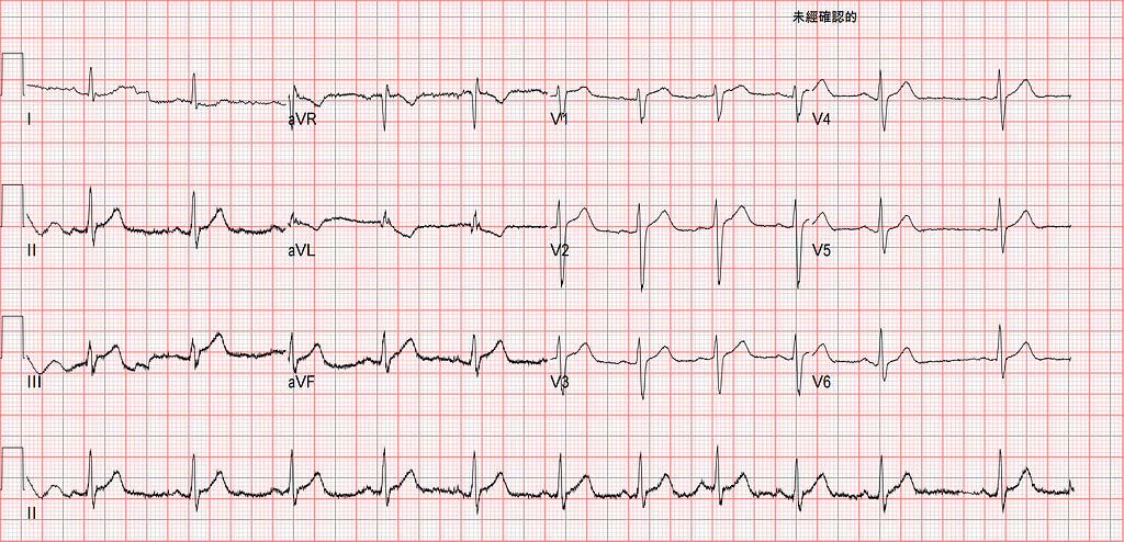
I remember this case was in April this year, one morning just as the COVID (Wuhan pneumonia, as it's often called in Taiwan) outbreak was taking off.
Back then, every day I was either at the quarantine facility, or at hotels screening migrant workers, or on my way to an ED shift.
That morning I was handed off a pulmonary edema patient who suddenly couldn't breathe, had chest pain, and whose blood pressure was down to the 50s. Then came a pile of trauma patients: facial bone fractures, plus vomiting blood. Another trauma patient had intra-abdominal bleeding on ultrasound; I shouted that this one was bleeding internally, and luckily a sharp team of nurses swarmed the patient, lines in, lines in, blood in, blood in. Waiting for arterial embolization to plug up the ruptured spleen.
After that came an OHCA (out-of-hospital cardiac arrest).
And it wasn't over. In came a 40-year-old man with typical chest pain, cold sweats, pain radiating to the left arm. While the ECG was being done, I said, no way it's going to be a STEMI right now, right?
Enough talk. Let's read this ECG.
Going through Rate-Rhythm-Axis-Interval-Ischemia, what are the findings?
Rate: about 66 bpm
Rhythm: P waves upright in I/II/aVF, inverted in aVR, and the P wave in II is taller than in I, so we can be sure the rhythm comes from the sinus node. Not perfectly regular; this counts as sinus arrhythmia.
Axis: Normal axis
Interval: no obvious QT prolongation
Ischemia: minimal STE in the inf.leads, plus obvious TWI in aVL.
Let's first ask ourselves a few questions; these will let us say with more confidence that this is an AMI.
Q1: Before STE shows up, are there any ECG ischemia signs that appear first? (Which ones are early ischemia signs?)
Q2: What kind of TWI in aVL can be normal?
Q3: Fig.1 is the ECG on arrival. If the chest pain continues, what ECG changes would we expect next? And if the pain goes away, what changes would we see?
We've always said that if you see a hyperacute T wave on the ECG, watch out, because ST elevation will start to show up next. In other words, the hyperacute T wave (HATW) may be an ischemia sign that appears even earlier than STE.
There's currently no firm definition of HATW. The great Smith describes the features of HATW → not tall, and usually not tall, but bulky and broad-based; the shape of the T wave has to be assessed together with the QRS height and the AUC.

The shape of Mount Fuji is a good way to describe HATW. Broad, bulky. And now I want to go to Japan again (click here)
Amal Mattu also often says that if you can fit the QRS inside the T wave, that T wave is abnormal.

So what exactly is the difference between the peaked T wave of hyper-K and the HATW of ACS?
Fig.2 is from the teaching images on Smith's ECG Blog: source here
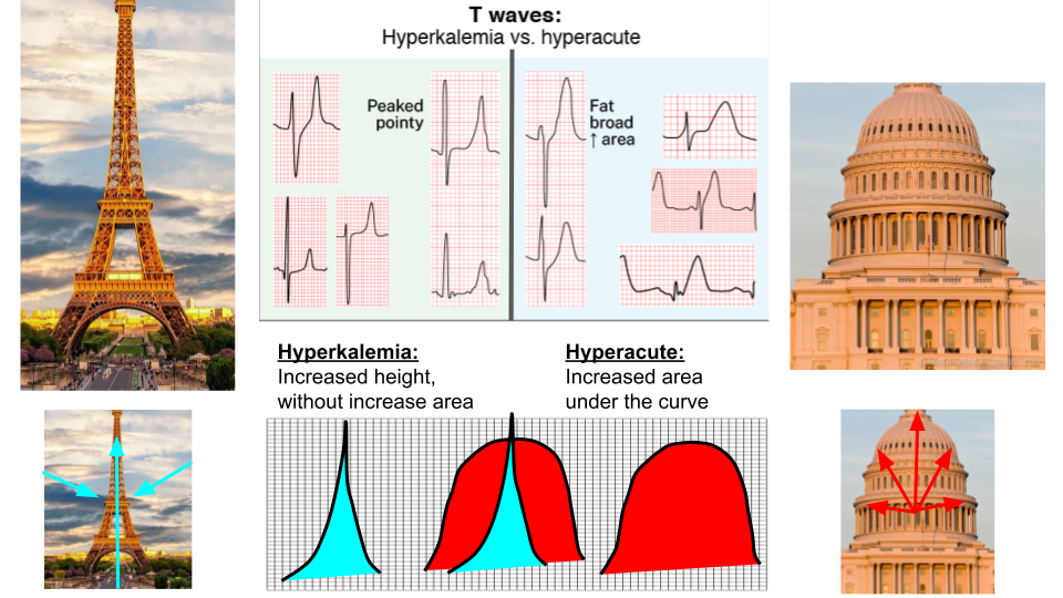
In Fig.2 you can see that the HATW T wave looks fat and bulky, and the AUC is increased (red area). The peaked T wave of hyper-K, on the other hand, has no increase in AUC but an increase in height, with a pointed tip, whereas the tip of an HATW is more blunt.
So in this case, the T waves in the inf.leads are actually relatively large, consistent with HATW.
Another key point is that this case has TWI in aVL. This has been emphasized in recent years as a possible early ischemia sign of an inf. wall STEMI.
In Aslanger's paper Ten steps to or away from cath lab1, step seven says to pay attention to subtle ECG findings (Fig.3) and look for any subtle reciprocal change.
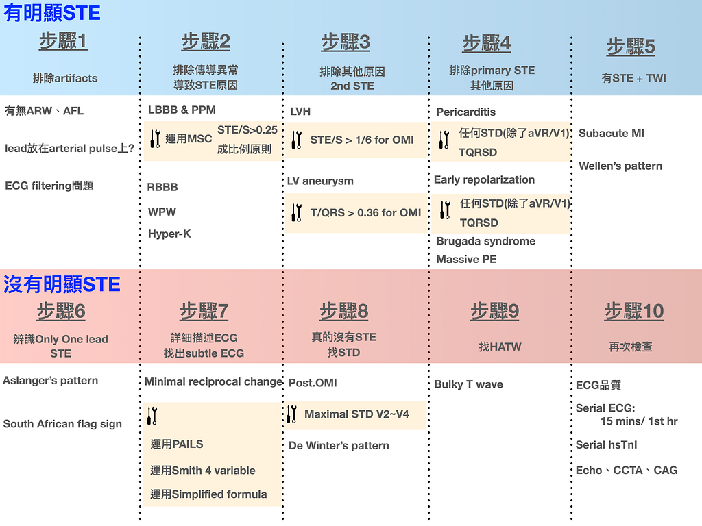
TWI in aVL (or STD in aVL) is the reciprocal change of ischemic changes in the inf.leads (especially Lead III).
Why does aVL mirror III? From Fig.4 you can see that I/II/III/aVR/aVL/aVF all sit on the frontal plane. III and aVL aren't exactly 180 degrees opposite, but they're still 150 degrees apart. So III/aVL are the pair of leads that best reflect each other's ischemic changes.

There's a key point to understand here: reciprocal change only appears in leads that face each other on the same plane.
Why stress this? Because there's a common misconception: when there's an ant. wall STEMI together with STD in the inf.leads, we're used to treating the inf. lead STD as the reciprocal change of the ant. lead STE. That's wrong!!!!!
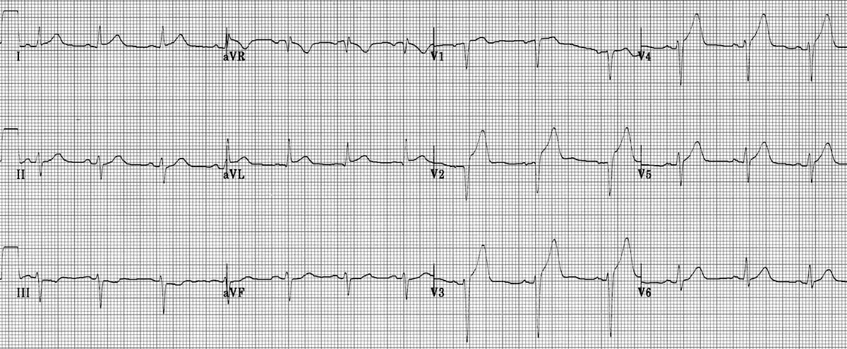
An example makes it clearer. See Fig.5.
Fig.5 is an obvious ant. wall STEMI with reciprocal STD in the inf.leads. So this is a true, genuine AMI.
The reciprocal STD in the inf.leads doesn't come from the STE in the ant. leads; it comes from the STE in the high lateral leads (I/aVL). I/aVL and the inf.leads belong to the same frontal plane. So the inf. lead STD here reflects that the high lateral leads are also involved in the AMI, which indirectly tells you the culprit lesion is highly suspected to be the proximal LAD.
Taking it further: which other leads on the ECG face each other like this and can reflect each other's ischemic changes?
- V1 and V6 sometimes show reciprocal change with each other → in a septal STEMI there's usually STD in V5-V6, because it's reciprocal to V1
- V7–9 (posterior leads) and V1–3 are leads that face each other
OK, I've wandered way off topic again. Back to the point: HATW and TWI in aVL can both be considered early ischemia signs. That is, signs that may show up before STE does.
Another ECG master, Jerry W. Jones, talked about the concept of reciprocal change in one of his lectures; I've put a screenshot of the slide below (Fig.6):
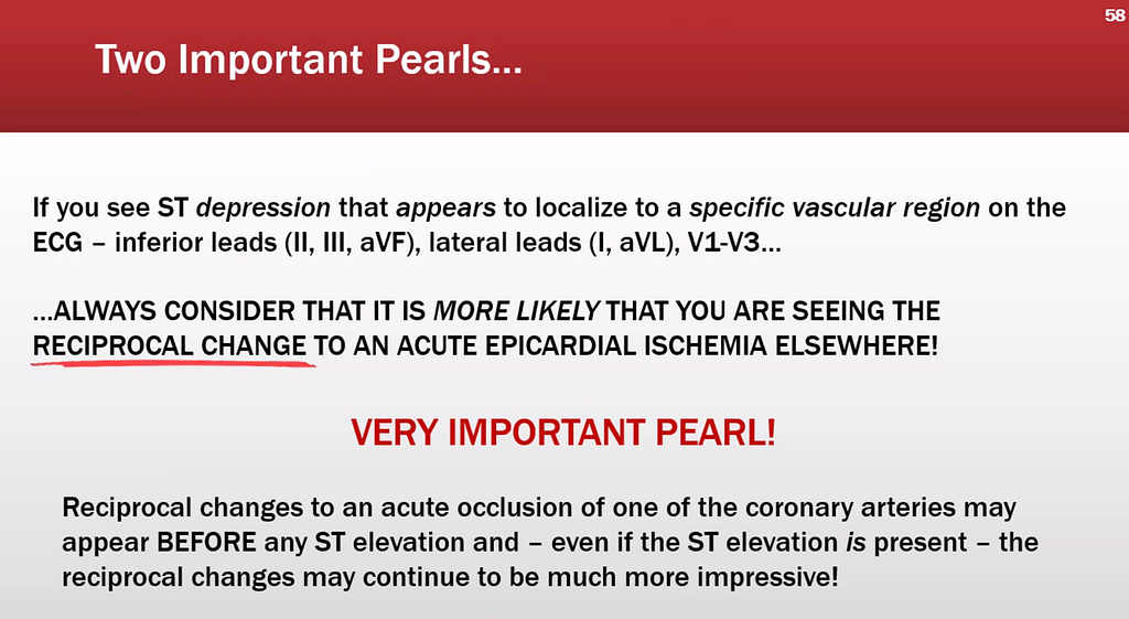
This slide (Fig.6) says that if the STD you see falls within a coronary artery territory, you must first think of this STD as the reciprocal change of STE on the opposite side, and quickly turn your eyes to the opposite side to see whether STE is there (be careful with any subtle STE)
The slide also says this reciprocal change may appear before any STE does. And if STE appears on the opposite side, the reciprocal STD may become even more obvious.
So when you see an early ischemia sign, remember not to park the patient somewhere way off at the far end of the ED. Put the patient where you can see them just by lifting your head, and if you also put them on an ECG monitor, that's the standard opening move every ED veteran has. Because in the blink of an eye, the ST segment may take off.
So what proportion of inf. wall STEMIs (OMIs) and ant. wall STEMIs (OMIs) show this reciprocal change?
👉 About 50-70% of ant. wall STEMIs have reciprocal change, which means 30–50% won't show reciprocal change (if I/aVL aren't involved, it won't be there) → so in ant. OMI patients you should not rely on the presence or absence of reciprocal change to rule out OMI.
- An ant. wall STEMI doesn't necessarily show reciprocal change over the inf.leads, especially when the occlusion isn't proximal.
- In an anterior MI, if there's STD in the inf.leads, i.e. reciprocal STD, it suggests the proximal LAD may be the problem (because only when the high lateral leads are involved do you see STD in the inf.leads). Mid/distal LAD usually doesn't produce reciprocal change
👉 Close to 99-100% of inf. wall STEMIs have reciprocal change (meaning in aVL)2
- So when you see "meaningful" TWI in aVL → wind yourself up right away and get ready to fight an AMI
- Note that an inf. wall STEMI without reciprocal STD over aVL doesn't mean you can rule out LAD occlusion (wraparound LAD)3
Now for question 2. Does TWI in aVL always mean trouble?
Amal Mattu has said in many ECG teaching sessions that to judge whether TWI in aVL is a problem, you need to pay attention to two things:
- TWI in aVL with a positive QRS (+) is the more meaningful one (a wide QRS-T angle is more likely an ischemia-induced STTC)
- LBBB and LVH both cause TWI in aVL (they're normal variants) → be careful to exclude these two first before analyzing further (that's what appropriate discordance means)
Also, the Aslanger paper mentioned earlier1 says that when looking over the whole 12-lead ECG for reciprocal change, be especially careful: LBBB, LVH, WPW, inferior LV aneurysm, and PPM usually have reciprocal STD at baseline with no ischemia.
Case continue:
The patient's symptoms were typical (chest pain, diaphoresis), and the ECG showed minimal STE over the inf.leads plus obvious TWI in aVL. The T-QRS angle of this TWI in aVL is wide (QRS positive, T wave negative), so this TWI in aVL is meaningful, and the patient had no LBBB or LVH.
I had the nurse practitioner call the CV man directly. The CV man on call that day didn't see a problem on the ECG, but said that since there were symptoms, they could still activate the cath lab and go in to see whether the vessels had a problem.
I had originally expected that with ongoing chest pain, the inf.leads would start to show STE any minute. (Note: TWI in aVL is an early sign of an inf. wall STEMI)
About 13 minutes later the patient felt a bit better, but still had pain (Fig.7).

After 40 minutes the patient felt much better (Fig.8).
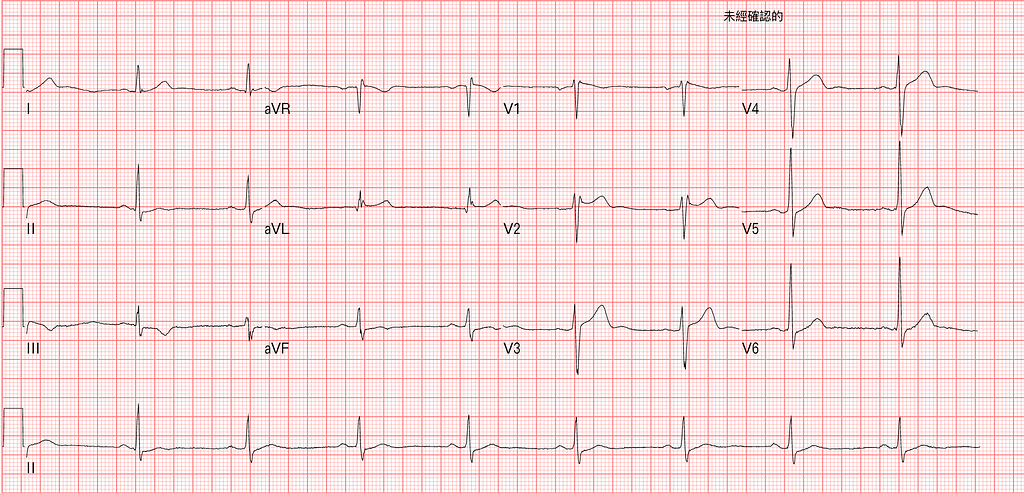
In Fig.8 you can see that as the symptoms resolved, a reperfusion rhythm appeared. Terminal TWI in II/aVF, and TWI in III. Because the patient had spontaneous reperfusion (the clot dissolved on its own and the vessel opened), you see these reperfusion T waves in the inf.leads, and the TWI in aVL flipped back upright. So this ECG means the vessel supplying the inf.leads had a problem, but it's open for now.
Does that last paragraph sound like something you've read somewhere before?
Yes, this is Wellens' waves showing up in the inf.leads. Wellens' waves don't only appear in the precordial leads; they can appear in the inf.leads too. What Wellens' waves fundamentally mean is that the vessel was occluded, but opened up before causing severe myocardial cell damage, so the leads supplied by that vessel show Wellens' waves.
As soon as the Wellens' waves appeared, I told the medical students on rotation that the ECG on arrival really did mean the patient had an occluded vessel. Maybe not an obvious STEMI, but definitely an OMI (Occlusion MI).
There were no beds that day, so the patient was transferred to another hospital for treatment. Later I followed up the transfer reply form for this patient. He had a cath at the other hospital.
CAD with SVD s/p PTCA+Stent
- Proximal RCA 90% stenosis
Learning points:
- Which leads on a 12-lead ECG may show corresponding reciprocal changes
- Reciprocal change caused by ACO (acute coronary occlusion) (e.g., TWI in aVL) may appear before STE shows up on the opposite side.
- Close to 100% of inf. wall STEMIs show reciprocal change in aVL, while 50–70% of ant. wall STEMIs show reciprocal change in the inf.leads, depending on whether the high lateral leads are affected. So if an inf. wall STEMI has no reciprocal change in aVL, you may need to think harder about whether something else is going on.
- What kind of TWI in aVL is meaningful? Which normal variants need to be excluded?
- What do Wellens' waves mean?
Department of Cardiology, Marmara University Pendik Training and Research Hospital, Istanbul, Turkey, Aslanger, E. K., Meyers, H. P., Department of Emergency Medicine, Carolinas Medical Center, Charlotte, North Carolina, USA, Smith, S. W., & Department of Emergency Medicine, University of Minnesota Hennepin Healthcare, Minneapolis, Minnesota, USA. (2021). Recognizing electrocardiographically subtle occlusion myocardial infarction and differentiating it from mimics: Ten steps to or away from cath lab. __Turk Kardiyoloji Dernegi Arsivi-Archives of the Turkish Society of Cardiology__, __49__(6), 488–500. https://doi.org/10.5543/tkda.2021.21026 ↩︎ ↩︎ ↩︎
Bischof, J. E., Worrall, C., Thompson, P., Marti, D., & Smith, S. W. (2016). ST depression in lead aVL differentiates inferior ST-elevation myocardial infarction from pericarditis. __The American Journal of Emergency Medicine__, __34__(2), 149–154. https://doi.org/10.1016/j.ajem.2015.09.035 ↩︎ ↩︎
You Diagnose Pericarditis at your Peril (at the Patient’s Peril!) ↩︎ ↩︎



Thoughts on this case? Leave a comment, a like, or a reaction below.
comments powered by Disqus