Translated from the original Traditional Chinese post. Read the Chinese original →
Chest Tightness That Started 2 Hours Ago: Is It AMI?
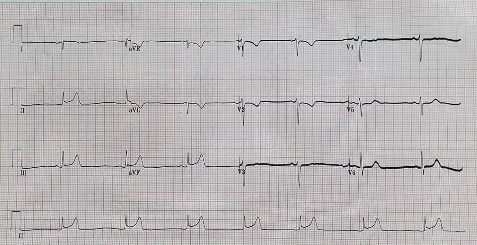
A 56-year-old man with no significant past history. In the small hours of the night he had chest tightness and discomfort, radiating to both arms.
Before handing off to me, my colleague asked whether this one had a problem.
I took a quick look on the spot. As I recall, I said: SB, minimal STE over inf.leads, TWI over aVL but no STD seen, STE over I, STD over V1~V2 with TWI
TWI in aVL does make you consider an early sign of inf.wall STEMI, but here the QRS-T angle is narrow, QRS(-), T wave(-). If the QRS-T angle here were wide, I'd find it more convincing. But if it isn't AMI, there seems to be no way to explain the STD over V1~V2 (Post.MI must be ruled out)
What looks like AMI:
- The symptoms fit
- minimal STE over inf.leads (must consider inf.wall STEMI)
- STD over V1~V2 (must consider inf.wall STEMI with post.involvement)
What doesn't look much like AMI:
- Narrow QRS-T angle: the QRS points in the same direction as the T wave, so the TWI may not necessarily mean anything
That's what I quickly assessed on the spot.
First, a few questions?
What rhythm is this ECG?
It's 3:30 in the morning and you're looking at this ECG. Would you Call CV?
If you didn't Call CV after seeing the first ECG, what should you do next?
First, let's answer what rhythm this is~~
Normally, if the SA node fires and conducts downward, the P wave is upright in Lead I, II, aVF and inverted in aVR
So what do we see on this ECG?
In Lead I the P wave is negative, the QRS is negative, and the T wave is negative. In aVR the P wave is positive. So it's not conducting down from the SA node? Or is it some other problem?
Tips: If you see RAD + an inverted P wave in Lead I, think of two DDx →Dextrocardia and lead misplacement

So how do you tell the two apart?
The more common lead misplacement is RA and LA swapped →you'll see aVR and aVL swapped, and the P/QRS/T in Lead I will be negative
Also, with lead misplacement, there's still R wave progression in the precordial leads
So let's look at the patient's arrival ECG again:
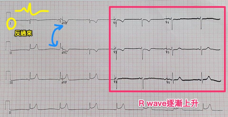
In the Precordial leads the R wave does gradually increase (at least it isn't gradually decreasing)
Swap aVL and aVR, and flip lead I over too—doesn't it look a bit more normal?
So this arrival ECG is still sinus rhythm (bradycardia), and the real aVL is actually the one labeled aVR (with STD + TWI)
Why does RA/LA reversal mislead us so easily here?
Because close to 99% of Inf.lead STEMIs show reciprocal STD change in aVL (in very early MI you may still not see it, and may only see TWI)1, so when we see STD in aVL, we can say with more confidence that this is AMI.
Tips: So in ant.wall STEMI, what proportion don't show reciprocal STD change? About 30~50%
Once aVR/aVL are swapped back, the original aVL with STD + TWI (i.e., the lead labeled aVR on the ECG) →turns into just TWI, no STD. That might make us lower our guard.
Next, what could TWI in aVL mean?
But if you see TWI in aVL, you still have to be very careful, because TWI in aVL can be a very early ischemia sign of inf.wall STEMI. You can't be too careful.
The same paper1 mentions that isolated TWI in aVL is 100% sensitive and 86% specific for inf.wall STEMI →in plain words, inf.wall STEMI almost 100% of the time shows TWI in aVL, so when it shows up, be careful—but it's not necessarily inf.wall STEMI; it could also be an LAD-m problem2.
Is TWI in aVL a normal variant or abnormal?
So when we see TWI in aVL, we need to worry about what comes next: will STE start appearing in the inf.leads? (Per the AHA NSTE-ACS guideline, do an ECG every 15~30 minutes in the first hour3).
At this point we need to ask an important question first: is this TWI in aVL normal (i.e., a normal variant) or truly abnormal?
You can use the QRS-T angle to assess further. The QRS-T Angle helps distinguish primary repolarization and secondary repolarization abnormality (note: these are abnormalities of repolarization). This Smith ECG blog post lays it out very clearly, and in it Ken Grauer explains a simple approach to the QRS-T angle4.
Key points written here:
- When TWI comes with any QRS-T Angle with an absolute value >45 degrees (calculate the QRS axis and the T wave axis and subtract)→you must think abnormal➡if it's abnormal, check whether the T wave in lead III is hyperacute, and you can also check for T wave flattening in V2 (which may mean a posterior OMI)
Ken Grauer's simple approach to the QRS-T Angle:
- What we're worried about is TWI over aVL →it may be normal, or reciprocal change STD from an inferior OMI
- That is, we don't care what the T wave axis of the T wave in aVL is (because we're only worried about an inverted T wave over aVL)(in other words, don't bother calculating the T wave axis)
- If the TWI in aVL is a normal variant➡you should see an rS-type QRS complex (clearly negative) in aVL. If you don't see a clearly negative QRS in aVL (or the QRS is positive), then the TWI in aVL is unlikely to be a normal variant (it may be from ischemia or infarction)
I've simplified the above into the flowchart below (Fig 1).

On with the case!!!!
In this case, the QRS in the real aVL was actually (-), and that really did lower my guard!!!!!
But the patient's symptoms persisted. 20 minutes later, I repeated the ECG (Fig.2)~~
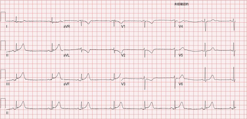
On this ECG, the electrodes are in the right places. You can see the STD in aVL is more obvious. And the STD originally in V1~V2 now extends to V3. There's definitely Post.wall involvement. The Hyperacute T wave in Lead III has become obvious. At that point you can diagnose inf.STEMI with post.wall involvement outright.
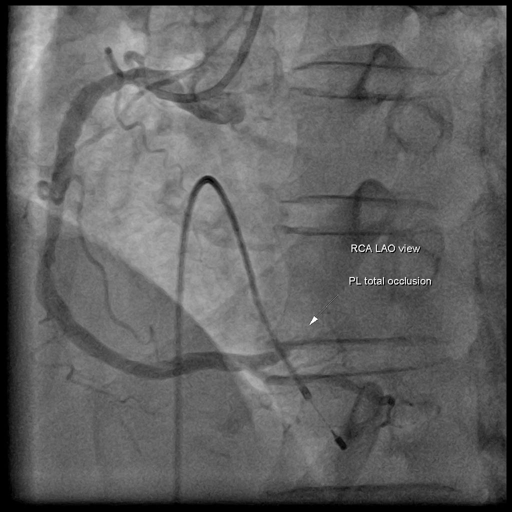
So 20 minutes after arrival, I called CV, and CV agreed with my diagnosis. The patient went straight to the cath lab. CAG report: RCA-PL total occlusion
Postscript:
- Honestly, the patient's initial QRS-T angle(-) with TWI really did lower my suspicion for AMI, but the clinical symptoms still couldn't rule it out. So after the patient arrived, I went straight to a Heart echo to see how the Inf.wall was moving. On the first Heart echo, by eyeballing, it really was moving pretty well. Before the patient went up to the cath lab, I wasn't ready to let it go and did another one (about 50 minutes after arrival), and only then did I see the basal septum moving worse.
This also taught me that ischemia signs on the ECG show up far earlier than LV RWMA on Heart echo!!!!!!!
Tips1: How to recognize RA/LA reversal
Tips2: What TWI in aVL means
Tips3: QRS-T angle wide + TWI in aVL →rule out LBBB, LVH
Tip4: Ischemia signs on the ECG show up far earlier than LV RWMA on Heart echo
Additional references: 1 2 3 4
Bischof, J. E., Worrall, C., Thompson, P., Marti, D., & Smith, S. W. (2016). ST depression in lead aVL differentiates inferior ST-elevation myocardial infarction from pericarditis. The American Journal of Emergency Medicine, 34(2), 149–154. https://doi.org/10.1016/j.ajem.2015.09.035 ↩︎ ↩︎ ↩︎
Hassen, G. W., Costea, A., Smith, T., Carrazco, C., Hussein, H., Soroori-Rad, B., Vaidian, S., Seashore, J., Alderwish, E., Sun, W., Chen, A., Simmons, B., Usmani, S., Kalantari, H., & Fernaine, G. (2014). The Neglected Lead on Electrocardiogram: T Wave Inversion in Lead aVL, Nonspecific Finding or a Sign for Left Anterior Descending Artery Lesion? The Journal of Emergency Medicine, 46(2), 165–170. https://doi.org/10.1016/j.jemermed.2013.08.079 ↩︎ ↩︎
Amsterdam, E. A., Wenger, N. K., Brindis, R. G., Casey, D. E., Ganiats, T. G., Holmes, D. R., Jaffe, A. S., Jneid, H., Kelly, R. F., Kontos, M. C., Levine, G. N., Liebson, P. R., Mukherjee, D., Peterson, E. D., Sabatine, M. S., Smalling, R. W., & Zieman, S. J. (2014). 2014 AHA/ACC Guideline for the Management of Patients With Non–ST-Elevation Acute Coronary Syndromes. Journal of the American College of Cardiology, 64(24), e139–e228. https://doi.org/10.1016/j.jacc.2014.09.017 ↩︎ ↩︎
Dr. Smith’s ECG Blog: A female in her 60s who was lucky to get expert ECG interpretation — link ↩︎ ↩︎



Thoughts on this case? Leave a comment, a like, or a reaction below.
comments powered by Disqus