Translated from the original Traditional Chinese post. Read the Chinese original →
This Is a STEMI. Do You Believe It?
Let's look at a case from my night shift yesterday. A 50-year-old man who worked catching glass eels (baby eels).
After reading some reports, I learned that glass-eel fishing is actually that dangerous. I'd assumed it was just kind of cold (standing in seawater).
Yesterday was freezing, the bone-chilling kind of cold. Last night, in the dead of the small hours, I only saw two patients—everyone would rather hide under the covers and sleep. That gives you an idea of how cold it was. Getting off topic~~~
While catching glass eels, this patient suddenly felt short of breath, chest tightness, and numbness in both hands.
Seeing the triage chief complaint, I really thought it was a hyperventilation attack. That cold, lower body soaked in seawater, such exhausting work—it actually fit the triggers for hyperventilation pretty well XD (I was wrong)
The patient was wheeled from triage to my exam room in a wheelchair. Since the chief complaint was chest tightness and dyspnea, I asked the nurse to do an ECG for me first. While it was being done, because the patient's breathing was shallow and fast and both hands were numb, I asked the patient to slow down their breathing, since they might really be hyperventilating a bit.
Tips: Hyperventilation is usually the effect; you need to dig into what the cause is. Gastroenteritis, big emotional swings, pain, and so on can all be triggers. I've even seen hyperventilation caused by aortic dissection. When the ultrasound picked up a flap floating in the aorta, I was breaking out in a cold sweat just like the patient. (Good thing I didn't write the patient off as a Psy episode XD)
Case continue
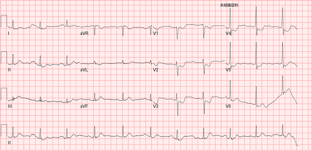
When the nurse finished the ECG, my first reaction was: this is a STEMI. Quick, move fast~~~~
I immediately wrote on the ECG: Post.MI
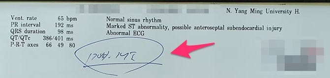
I asked the nurse practitioner to send it right away to the on-call CV man.
First, let's read the ECG.
Rate: 65
Rhythm: Sinus rhythm (P waves upright in Lead I, II, aVF, inverted in aVR)
Axis: Normal axis
PR slightly long, Narrow QRS, No QT prolong
STD in V1~V5, Maximal STD in V3~V4, no obvious STE
WTF~~~ No STE, and you're telling me this is a STEMI!!!!
Yes. This is a posterior STEMI, so you can't see any STE at all on the 12 leads ECG. But it truly is a STEMI.
Post.wall MI makes up roughly 10~20% of all STEMIs. And it usually comes together with inf.wall MI and Lateral wall MI.
Of all MIs that involve the Post.wall, 90% come with inf.wall and lateral wall involvement
Only 4~10% are isolated Post.wall MI.

Let me use an example to explain the sentences above; it'll make this clearer.
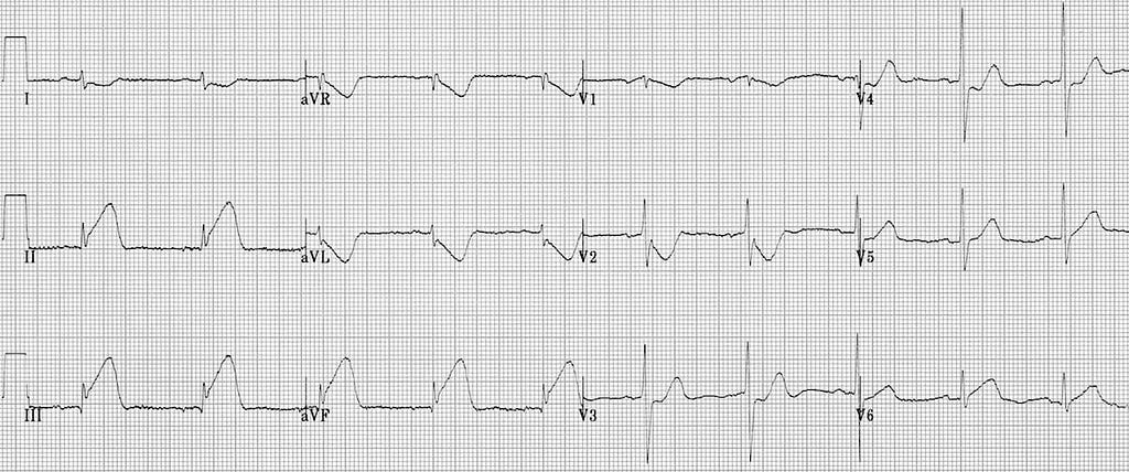
When a patient clinically has chest pain and diaphoresis and gets an ECG like Fig.1, we'd never miss it. STE in the Inf.leads as tall as mountains will definitely make us itch to consult the CV man. But what if this patient had no STE at all in the limb leads (left side of Fig.1), and only the precordial leads showed changes like Fig.1? Would you be able to say right away that this patient has an MI?
I think probably more than half of people would rather wait for the biomarker results and consult the CV man if they come back high.
The difficulty in diagnosing Post.wall MI is that we're all held hostage by the STEMI criteria. You have to see STE before you can consult the CV man. If it's not STE, then maybe it's NSTEMI? That's exactly where Post.wall MI most easily gets misdiagnosed.
In Post.wall MI only 30% of patients achieve D2B<90 minutes, and the reason is that, because we don't see STE and only see STD, we treat it as NSTEMI, which delays PCI.
So isolated Post.wall MI is even easier to misdiagnose, because there's no accompanying inf.wall or lateral wall MI. With no STE in those leads, it's even easier for us to overlook that an emergency is going on.
Tips: We easily misdiagnose Post.wall MI as NSTEMI, mainly because we're tied down by the concept of the STEMI criteria
Case continue
After the CV man looked at the ECG, the CV man asked us to do a set of Post.leads.
ECG fans who follow the Smith ECG Blog will know that Dr. Smith actually doesn't favor doing Post.leads to diagnose Post.wall MI.
Let me explain to you readers, and you'll see why he doesn't favor Post.leads: because it delays PCI.
Look at Fig.2. First, where do the Precordial leads sit on the body relative to the heart? These Precordial leads are almost right on the surface of the heart. But what about the Post.leads? Post.leads means doing V7~V9—that is, moving V4~V6 to V7~V9, placed on the back.
In Fig.2 you can see that if the cardiac impulse travels along the axis, to reach V7~V9 it runs into a big chunk of lung tissue. When we learned ECG, we also learned that when we see low voltage, we put COPD and hyperinflated lungs on the differential. Because the air separates the electrodes from the heart. That makes the QRS complex amplitude smaller. And when the QRS voltage gets smaller, the accompanying ST segment deviation and T wave shrink proportionally too.
In plainer words: if there's a Post.wall MI, most of the time you can see Post.lead STE. But—and it's this "but"—the voltage in the Post.leads is usually small, so the STE won't be obvious.
So you might misread it as no STE, when actually, if you apply proportionality and look at the ratio of STE to QRS, there really is ST elevation; it's just very subtle.
So if you want to use Post.leads to help diagnose Post.wall MI, one very important concept is: if you see STE, the probability of Post.wall MI is high (high Specificity), but if you don't see STE in V7~V9, that doesn't mean there's no Post.wall MI, because the low voltage makes visible STE in the Post.leads hard to see (Sensitivity isn't high).

So how high does visible STE have to be to count as meaningful?
- For both men and women, STE of ≧0.5 mm (in any lead) in V7~V9 counts as meaningfully elevated.
- In men <40 years old, seeing >1 mm of STE in V7~V9 (any lead) increases specificity.
Tips: No STE in the Post.leads doesn't mean the patient has no Post.wall MI
So when exactly should you suspect Post.wall MI?
When the Maximal STD is in V1~V4, you should strongly suspect it. Once there's STDmaxV1–4, it has 96% specificity—meaning no matter what, you must do everything you can to rule it out; otherwise this STD is a Post.wall MI.
Tips: STDmaxV1–4(+) →points straight to Post.wall MI
Case continue
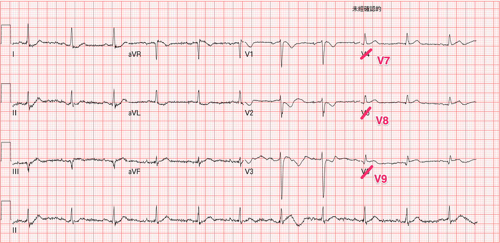
Fig.3 is the Post.leads ECG done in the ED. Let's blow up V9 for a closer look (Fig.4)
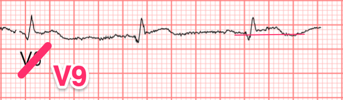
You can see at least 0.5 mm of STE in V9, which confirms this is a Post.wall MI
There's a very important concept here, also mentioned in the latest Smith ECG blog post1. That is: if we're going to do Post.leads, never move the V1~V3 electrodes to the back to make V7~V9.
Why?
Stephen Smith says that when doing Post.leads, never change V1~V3 into V7~V9. Because a coronary thrombus is dynamic and may spontaneously reperfuse; if you happen to do the post.leads during a reperfusion phase, then of course you won't see STE in V7~V9—so keep V1~V3 in place for comparison.
Tips: When doing Post.leads, don't move V1~V3 to make V7~V9
At the time I did an Echo with my own Lumify. Where would you look, and what problem would you hope to see?
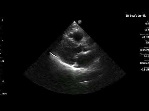
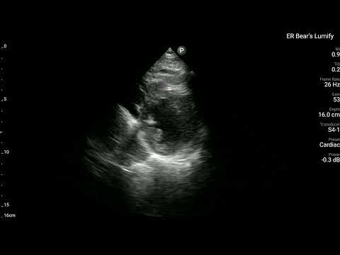
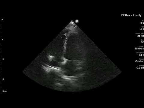
Under the 17 segment model, the LV is divided into 17 segments. The Post.leads location corresponds to the Basal-inferior area (Segment 4).
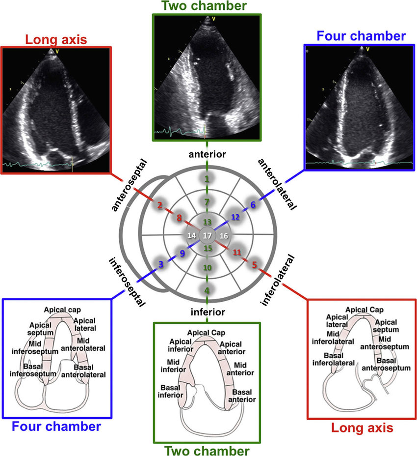

First let's look at the right side of Fig.5, where you can clearly see the 17 segment layout.
So when the ECG makes us suspect Post.wall MI, of course our ultrasound gaze goes to around the Post.wall (look at Segments 4, 5, 10, 11) for obvious RWMA. If there is, the ultrasound findings match the ECG findings perfectly, which further proves this really is a Post.wall MI.
Looking at the left side of Fig.5, you can tell that A4C actually doesn't show the Post.wall well. A2C and PSAX can.
- A2C looks at anterior and inferior, and can show Segment 4
- PLAX can show Segment 5
- PSAX looks at a cross-section (short axis), and can show Segments 4, 5, 10, 11
PSAX is the very best view for seeing the Post.wall and RWMA around it.
Go back to the POCUS Clip above (PSAX view), put a finger in the middle of the LV, and notice whether the area around Segments 4 and 5 isn't really moving.
Bingo!!!!
POCUS finding is compatible with ECG finding.
Tips: If you suspect Post.wall MI →PSAX view is the best view for eyeballing
Case continue
Of course, given all the Post.wall MI evidence the ED put together, an emergency cath was activated.
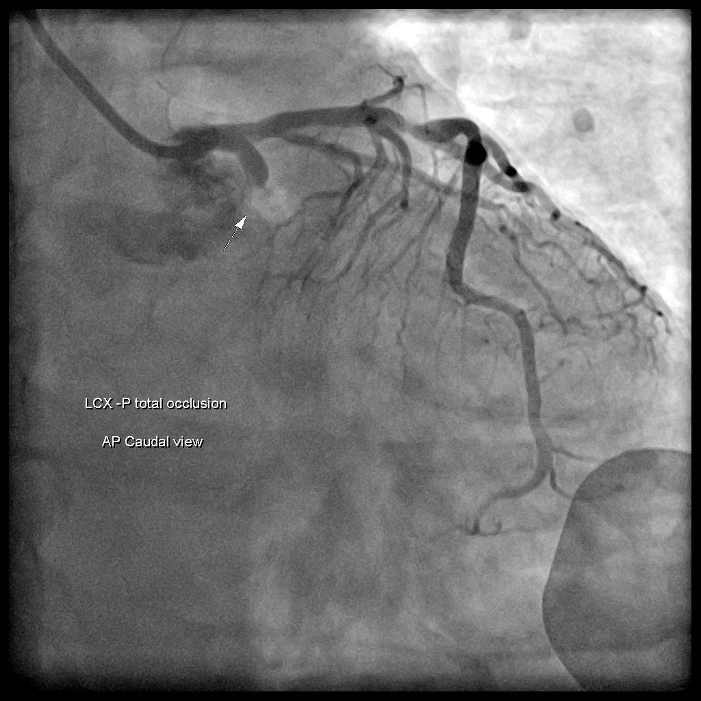
Seeing Isolated Post.wall MI➡it's more commonly LCx occlusion than RCA occlusion➡usually in these cases (isolated Post.wall MI) the PL branch can't supply enough blood to prevent ischemia (if it could, you wouldn't get isolated Post.wall MI).
CAG finding: LCx-P total occlusion
Learning Points:
- Hyperventilation is usually the effect; you need to dig into what the cause is. Gastroenteritis, big emotional swings, pain, and so on can all be triggers. I've even seen hyperventilation caused by aortic dissection. When the ultrasound picked up a flap floating in the aorta, I was breaking out in a cold sweat just like the patient. (Good thing I didn't write the patient off as a Psy episode XD)
- We easily misdiagnose Post.wall MI as NSTEMI, mainly because we're tied down by the concept of the STEMI criteria
- No STE in the Post.leads doesn't mean the patient has no Post.wall MI—don't waste time doing Post.leads
- STDmaxV1–4(+) →points straight to Post.wall MI, until ruled out
- When doing Post.leads, don't move V1~V3 to make V7~V9
- If you suspect Post.wall MI →PSAX view is the best view for eyeballing
Additional references: 1



Thoughts on this case? Leave a comment, a like, or a reaction below.
comments powered by Disqus