Translated from the original Traditional Chinese post. Read the Chinese original →
Heart Warning: Pericarditis vs. OMI — Readmitted
Let me tell you about a case a colleague ran into recently. I think there's a ton to learn from it. Let's go panning for gold in this one together~~~

The patient is a 39-year-old man with a history of hypertension, hyperlipidemia, and spontaneous intracerebral hemorrhage.
On his first visit, he told us in the ED that his chest felt tight, that it hurt, and that he had palpitations.
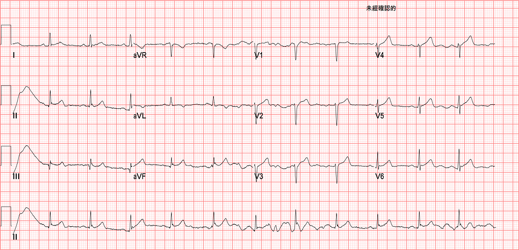
Fig.1 is the ECG from that first visit.
A quick read:
The baseline in II/III is unstable. That's because the LL electrode is shaking.

How do you tell within 5 seconds of looking at an ECG that it's a baseline artifact from a shaking left leg?
Picture Lead I/II/III in your head. The electrode that makes both II and III shake is the LL electrode.
For beginners reading ECGs, I recommend going in the order Rate-Rhythm-Axis-Interval-Ischemia. That way you're less likely to miss things or skip anything.
Rate: 70 bpm
Rhythm: SR (P waves upright in I/II/aVF, and the P wave inverted in aVR)
Axis: normal axis
For axis, I recommend playing around with this website. Spin the dial and watch how I/aVF/II change, and you'll get a feel for which way the axis is pointing~~~
Interval: No QT prolong
Ischemia: The first goal is to look for any OMI ECG findings (currently 18 of them… and the list is only going to get longer XD)
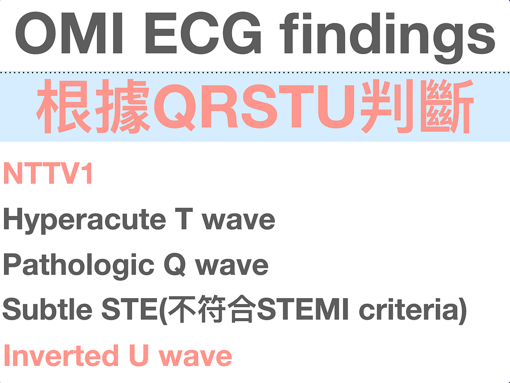
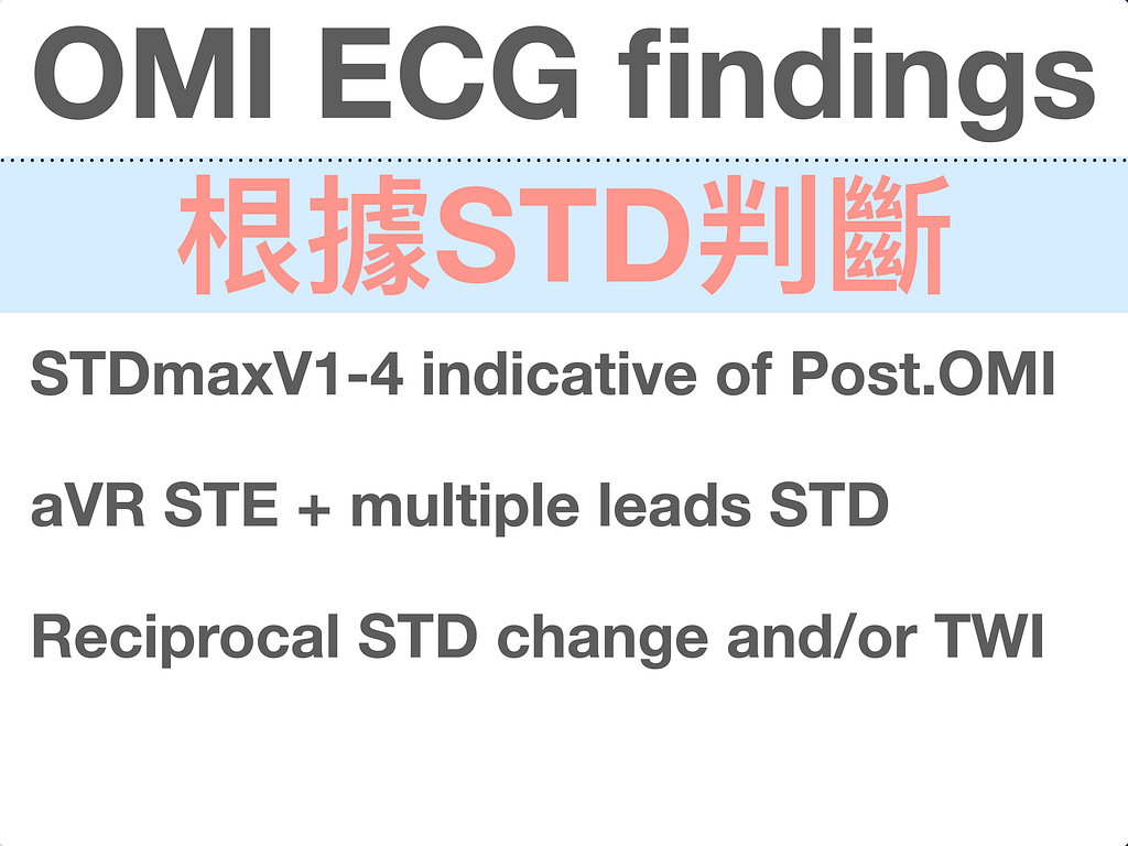
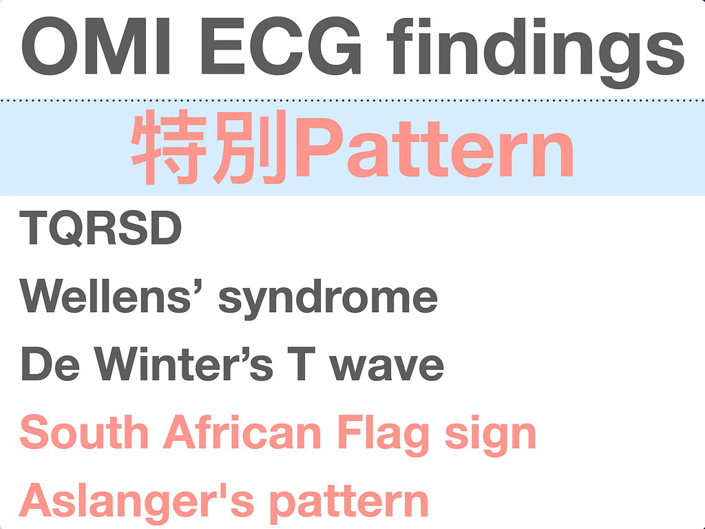
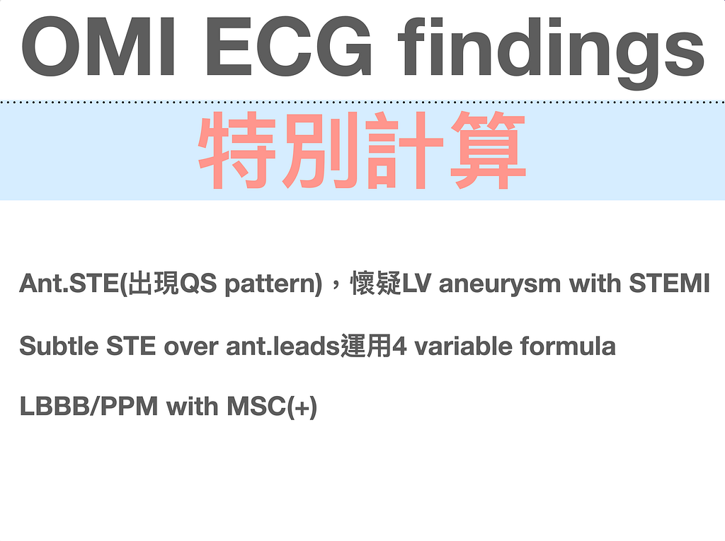
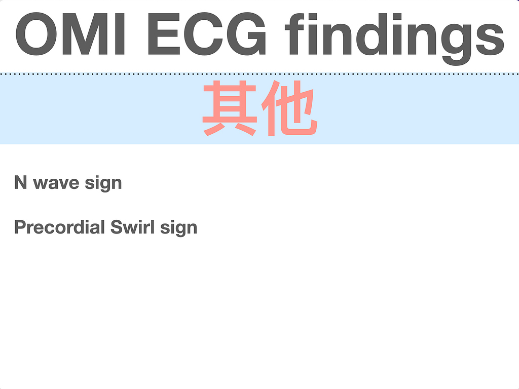
OMI(Occlusion MI)
If you read Dr. Smith's ECG Blog and Ken Grauer's ECG interpretation site, you probably know exactly what I'm talking about.
Those of us on the front line facing patients have to do everything we can to catch every OMI, and not send any of them home.
Of all the patients who come through the ED every day, a certain proportion always show up with ACS symptoms. We have to be confident we can catch every OMI.
If we don't catch it….. this is how the story usually goes.

An in-hospital call from a colleague…….
Hello~~ Dr. Bear? That patient you saw….. a few days ago…..
Before they even finish the sentence, I think anyone who works in the ED, hearing just those few words, gets peripheral hypoperfusion and cold extremities on the spot. That's a shock sign……. and there's no fix XD
Getting better at reading ECGs is, I think, the only answer~~~~
First let's look at what it actually means to catch an OMI patient. I think this is pretty important.
So what kind of OMI patients are we trying to catch?
STEMI(+)/OMI(+) and STEMI(-)/OMI(+)
STEMI(+)/OMI(+) needs no explanation. We should be catching those anyway, though you do have to watch out for STEMI mimics. (I'll write a blog post on that when I get the chance)
The ones we really need to catch are STEMI(-)/OMI(+). In plain words: these patients don't meet STEMI criteria, but the artery really, truly is occluded.
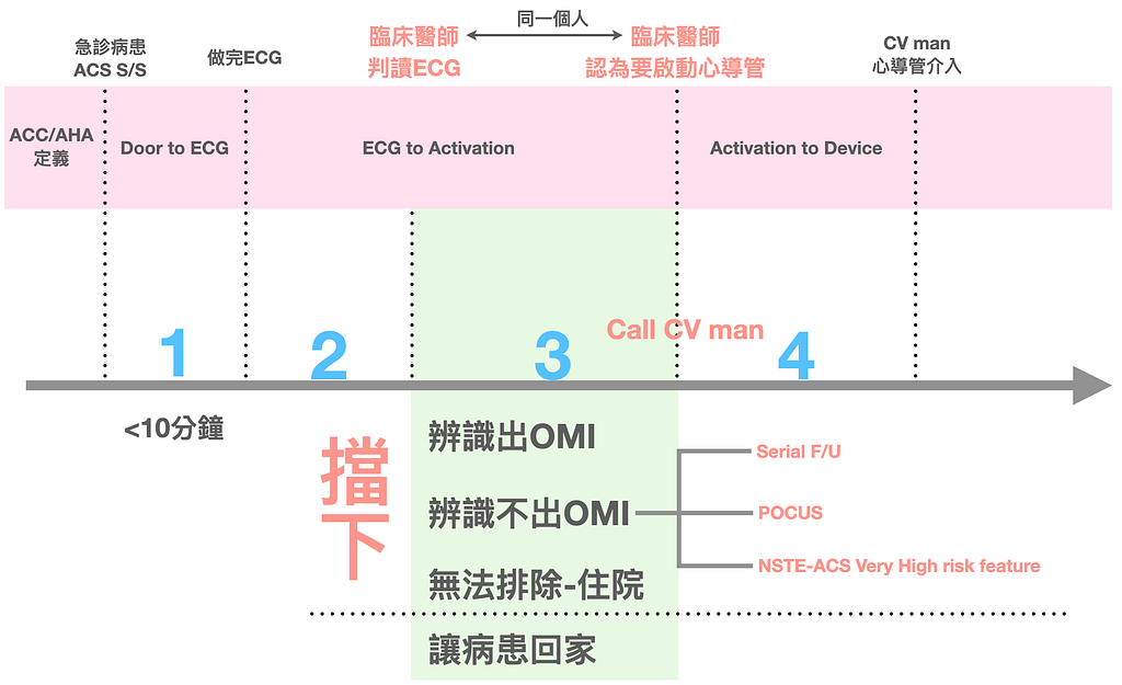
Catching works at several levels (see Fig.3)
Segment 1 in Fig.3 is something hospital accreditation already audits (and the guideline says <10 minutes too). Is there anyone who'd dare not get an ECG done within 10 minutes? (Door to ECG time)
Segment 2 runs from the ECG being done to the clinician reading it. This usually depends on the clinician, because once the ECG is done it should land on the physician's desk pretty quickly. Unless the physician doesn't see it, or doesn't realize there's a chest pain patient's chart sitting on the desk. (The time for 2 is almost negligible. If it's getting longer, look into why the ECG sat so long after being done without a clinician reading it)
Segment 3 I think is extremely, extremely important. This stretch runs from the clinician starting to read the ECG to the clinician deciding the cath lab should be activated. It's called ECG to Activation time (ETA). (Ps: note this is the same person. It's not the CV man deciding to activate the cath lab, it's the physician reading the ECG deciding to activate it)
Segment 4 is the time from calling the CV man to the catheter intervention (Activation to Device). Honestly, there's not much the ED man can do during this stretch, because it depends on whether the CV man thinks the patient needs PCI. We may think the patient needs Cath lab activation and call the CV man, but the CV man may think it's not needed.
The ACC/AHA D2B time includes all of the above, with a target D2B time of <90 minutes1.
Next, let's break down what catching means in Fig.3
- Recognize the OMI → this includes STEMI(+)/OMI(+) and STEMI(-)/OMI(+) → Call CV man
- Can't recognize the OMI → but know how to use the following tools and patient features → if (+), Call CV man
- Serial F/U ECG + Enzyme (after the initial ECG, you feel something's off with the ECG but it doesn't meet STEMI criteria, and you're not that familiar with the 18 signs of OMI ECG findings, but you just think the ECG has a problem: please, please remember F/U ECG within 10–15 minutes)
- Use POCUS to look for RWMA
- If the patient doesn't meet STEMI criteria but has a very high-risk feature of NSTE-ACS → Call CV man (I've brought this up many times in earlier blog posts, not repeating it again)
- ACS can't be clinically ruled out, so in the end we admit the patient
Being able to Call CV man or decide the patient should be admitted → that's catching it.
The worst scenario is not recognizing the OMI, not doing F/U ECG + Enzyme, and letting the patient go home.
Why I say the ETA time in Fig.3 is extremely, extremely important~~~~
The point of the ETA time in Fig.3 is that clinicians need to be good enough at reading ECGs to shorten the time from reading the ECG to calling the CV man.
ETA time can be shortened through training. This is the only way to improve ourselves and help patients. (Changing yourself is far more effective and useful than changing other people)
Here I recommend a paper McLaren published in AJEM in 20212.
The paper compares one year pre-intervention with one year post-intervention:
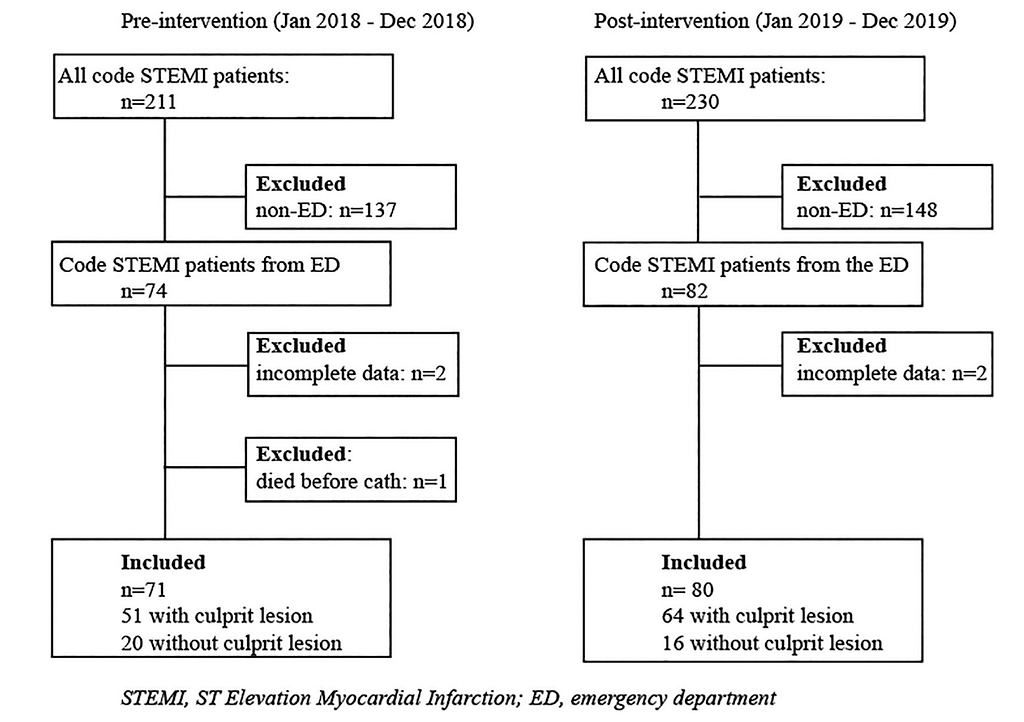
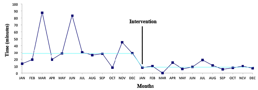
Fig.4 shows that the intervention shortened the ETA time.
So what did the intervention in the paper include?
According to the authors, it included grand round presentations, plus weekly posts on the internal network of challenging case ECGs with STEMI equivalents and subtle occlusion, along with literature discussion. They also tracked the number of views/visits to the case ECGs on the internal network.
The paper's conclusion was that after the intervention started, ETA time went down, without a significant increase in the number of Code STEMI w/o culprit lesion.
What this means is that the intervention taught the physicians the OMI ECG findings and shortened the time from the ED man reading the ECG to the ED man deciding the cath lab needs to be activated (Call CV man). That time saved didn't come from calling at random. These Code STEMIs (Call CV man) were all meaningful calls, because statistically there was no increase in the proportion of Code STEMI w/o culprit lesion.
The paper's appendix gives 10 cases of ECGs that look like they might be normal but were actually ACO patients. If you're interested, download the paper and read it yourselves~~~~
Back to Case
Without giving away yet what Fig.1 shows, the patient was later admitted to the cardiology ward, and troponins were drawn along the way.

During the admission, the patient had an Exercise stress test and an Echo.
According to the chart, the Exercise stress test was normal. As for the echo, I didn't see any report.
The patient's final discharge diagnosis was:
Chest pain, favor pericarditis related
First of all, what's really odd is that the troponin was already above the 99th percentile of URL on the initial ED draw, and it went even higher on days 2-3 on the ward. This is definitely not Chronic myocardial injury.
Also, if this really is pericarditis, we have to ask ourselves a question: can pericarditis come with a rise in biomarkers?
This UpToDate article3 describes a study of 118 patients diagnosed with acute idiopathic pericarditis, in which 38 patients (32%) had elevated biomarkers. But in these patients you should also consider whether it's myopericarditis. (Fewer than half of pericarditis cases have a biomarker rise)
Dr. Smith has a post on diagnosing pericarditis that is an absolute classic4.
That post describes how if the troponin is up, AMI vs. pericarditis turns into AMI vs. myocarditis.
And in the ED, AMI and myocarditis can't be told apart. It takes Cath lab activation to tell them apart.
In the comments on another post, the master, Ken Grauer, stresses one point even more5:
Even on international ECG forums, pericarditis is still very rare clinically → Smith's ECG Blog stresses this over and over: pericarditis is much rarer than OMI or a repolarization variant. Stephen Smith often says "You diagnose acute pericarditis at your peril!"
So what exactly does "You diagnose acute pericarditis at your peril!" mean? I asked ChatGPT to translate it, and I think it nailed it.

A wrong diagnosis can have serious consequences for the patient.
Yep~~~~ a whole bunch of masters have said it: diagnose pericarditis, and the consequences are on you!!!!!!
The consequences come from deciding it's pericarditis without ruling out the other problems (OMI or myocarditis).
Next, let's look again: this patient had an exercise stress test with a normal result. Does that count as ruling out OMI?
No (big font, saying it again and again: a normal exercise stress test cannot rule out a problem in the patient's arteries)
Why?
We have to ask a question. Is the Exercise stress test reliable?
From this article6, we learn a few key points about the Exercise stress test:
⭐️A negative exercise stress test is of no use whatsoever to us ED men
- Plenty of ED men have probably had the experience of treating quite a few real ACS or cardiac arrest patients, some of whom had a recent negative exercise stress test.
- Over-relying on a negative exercise stress test is a common reason patients get delayed or misdiagnosed.
Because we may imagine that a Negative exercise stress test = nothing wrong with the arteries (but that's not true)
- It's important to remember that coronary artery disease (CAD) can be caused by atherosclerotic lesions with only mild stenosis and unstable plaque rupture, which a standard exercise stress test can't detect.
Also, in the March 8, 2021 ECG weekly7, Amal mattu made a special point of one concept.
Right at the start of this lesson, Amal asks:
Why do so many AMI patients who recently had an exercise stress test or CAG showing no severe occlusion go on to have a real AMI just a few days later, and the CAG then finds a very tight occlusion?
Let me explain it step by step~~~
Whether it will lead to ACS → stress tests and Cath don't help all that much → this can be explained clearly by the pathophysiology of ACS
How much plaque vulnerability (how easily it gets damaged) and stability matter in causing ACS >> plaque size and the degree of luminal stenosis
Whether a plaque is vulnerable depends on three key factors:
- The size of the Fibrous cap
- How much Lipid core there is
- Inflammatory cells
1️⃣ The problem with the Exercise stress test
- Smaller plaques are more likely to be unstable and rupture
- These small plaques can still rupture and cause complete occlusion → studies have shown that before the rupture that causes AMI, the IRA (infarct related arteries) usually had no occlusive plaque (because the plaque was small, it wasn't occluding)
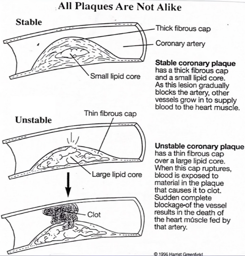
Fig.5 shows that not all plaques are the same
- Stable coronary plaque → thicker fibrous cap, less lipid core (in this figure the fibrous cap is thick enough to narrow the lumen, so a stress test will come back positive
- Unstable coronary plaque → thinner fibrous cap, more lipid core (in this figure the lipid core + fibrous cap may not even block half the lumen, so a stress test may come back negative, but its composition is unstable, which makes it an unstable plaque) → when the fibrous cap ruptures, blood and the lipid core form a clot, which then occludes the artery
2️⃣ The problem with Cath
- It can't tell stable vs. unstable plaque composition apart → because Cath can't tell how much fibrous cap and lipid core there is
- It can't detect "coronary artery remodeling"
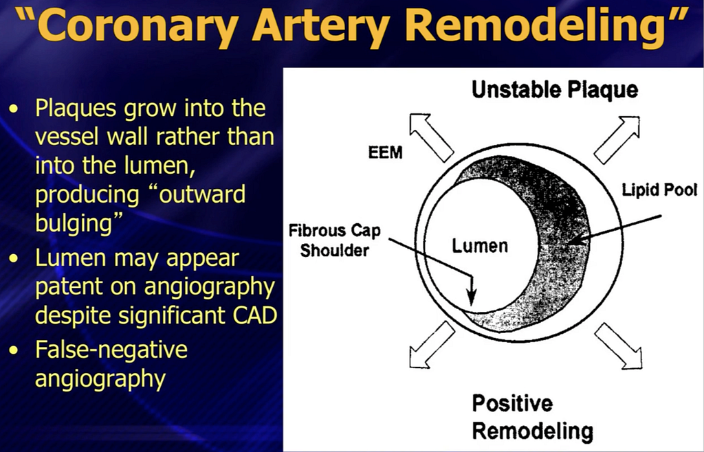
- Fig.6 shows the Plaque growing into the vessel wall (outward bulging) > growing into the lumen → so on CAG the artery may look patent, but in fact significant CAD is already there → which is how you get false-negative angiography
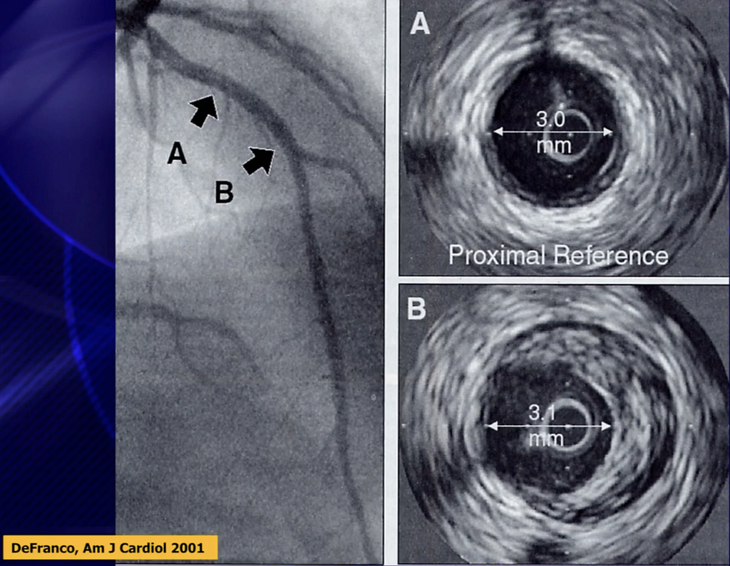
- Fig.7 shows that on CAG, A and B both look almost entirely open (about the same luminal diameter), but when you actually look with intravascular US (IVUS), there's a large plaque at B. Because of remodeling the lumen still looks big enough, but the rupture risk is actually high.
- B is more stenotic than A (without IVUS, you'd just think B looked open, but in fact it's already stenotic)
Putting all of this together: even if a recent stress test or CAG was Negative, it can't completely rule out ACS (no single workup can bring the risk of ACS down to zero) → the solution: history matters, ECG, TnI, a validated ADP (HEART score, etc.)
- Of course, if the CAG report says clean coronary artery, the chance of ACS from plaque rupture is low. But if the report says insignificant or unremarkable CAD (since CAG can't tell plaque vulnerability and stability), be careful
Back to Case, Again
On the morning of day 4 after being discharged with a diagnosis of pericarditis, the patient suddenly felt waves of tight, bloated pain in the left chest, with pain in the left upper back too. He said he'd been fine since discharge, the pain only started today, and it felt the same as during his last admission.
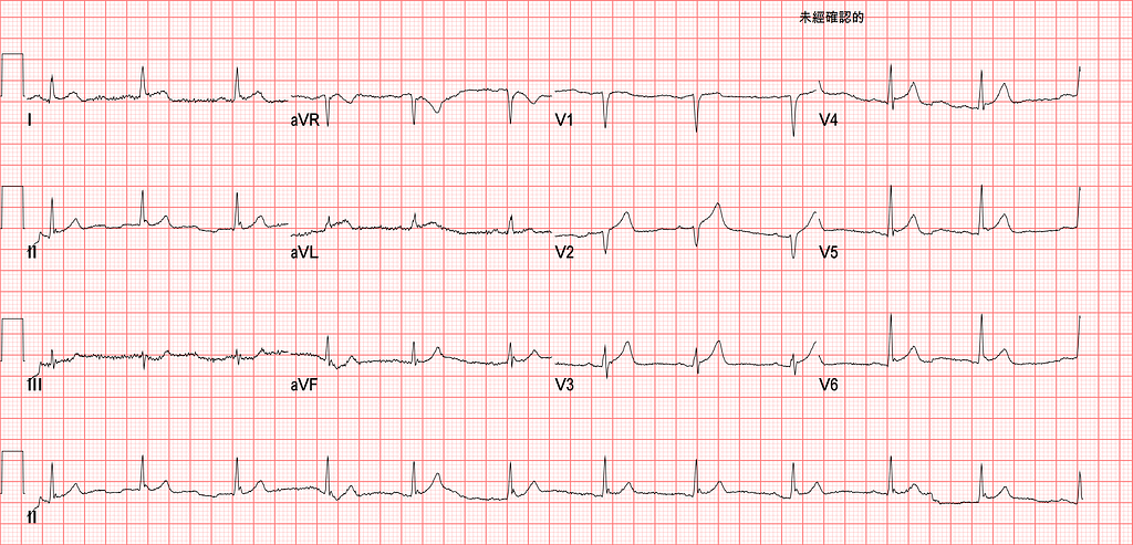
What ischemic ECG morphology do you see in the Fig.8 ECG?
- First, V1-V4 have LV aneurysm morphology, so we can apply Dr. Smith's T/QRS >0.36 → V2/V3 definitely exceed 0.36
- Straight ST segment over I/aVL, Maybe STD in III
- Is the STE in V4-V6 meaningful? I think I see J waves, so to me it looks more like STE from early repolarization
Putting it all together, we can infer the problem is probably in the proximal LAD. It also seems to fit the South African Flag sign → suspect D1 occlusion
If we go back to Fig.1 (the initial ECG from the first ED visit), we can see V1-V4 also fit the LV aneurysm pattern, so T/QRS > 0.36 applies, and if any one lead meets it, you can diagnose STEMI → V4 in Fig.1 is absolutely >0.36
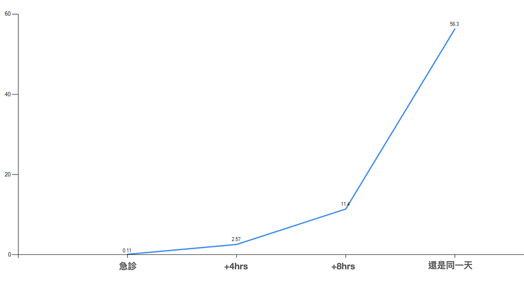
Later that same day, the patient's troponin came back sky-high…….
But the CV man still didn't schedule a CAG that day (even though it was a weekday, during the day)
Hmm…….. this is exactly what I meant about segment 4 in Fig.3: there's nothing we ED men can change about it. Because it depends on the CV man's attitude toward this Case.
CAG final result: Acute D1 total occlusion
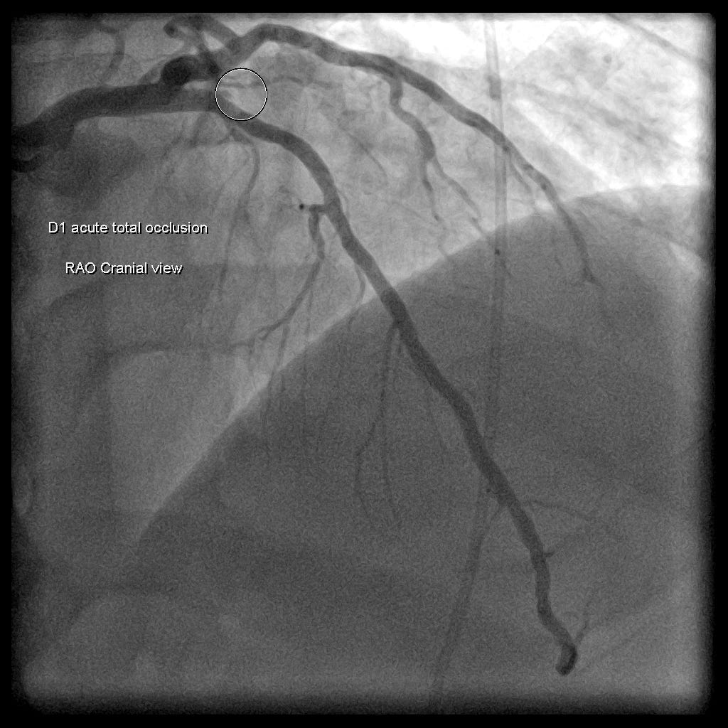
Quick wrap-up of this case
First of all, the first visit was probably already an OMI (T/QRS>0.36 in V4, and the biomarker was up)
But just as Dr. Smith says:
You diagnose acute pericarditis at your peril!
When we're about to make the diagnosis of pericarditis, we have to do everything we can to rule out OMI.
And if we don't understand what a Negative exercise stress test means and the principle behind it, we'll be blindfolded and leave the patient on the edge of danger without even knowing it!!!! Seriously!!!!
Key points:
- What are the 18 OMI ECG findings?
- How to catch OMI patients
- Ways to reduce ETA
- Do we really want to diagnose pericarditis?
- Can a recent Negative Exercise stress test or CAG rule out ACS?
- When the ECG morphology fits LV aneurysm, you can apply T/QRS >0.36 (I covered this in a few earlier blog posts, have a look)
Further references: 1 2 3 4 5 6 7
O’Gara, P. T., Kushner, F. G., Ascheim, D. D., Casey, D. E., Chung, M. K., de Lemos, J. A., Ettinger, S. M., Fang, J. C., Fesmire, F. M., Franklin, B. A., Granger, C. B., Krumholz, H. M., Linderbaum, J. A., Morrow, D. A., Newby, L. K., Ornato, J. P., Ou, N., Radford, M. J., Tamis-Holland, J. E., … Zhao, D. X. (2013). 2013 ACCF/AHA Guideline for the Management of ST-Elevation Myocardial Infarction. Journal of the American College of Cardiology, 61(4), e78–e140. https://doi.org/10.1016/j.jacc.2012.11.019 ↩︎ ↩︎
McLaren, J. T. T., Taher, A. K., Kapoor, M., Yi, S. L., & Chartier, L. B. (2021). Sharing and Teaching Electrocardiograms to Minimize Infarction (STEMI): reducing diagnostic time for acute coronary occlusion in the emergency department. The American Journal of Emergency Medicine, 48, 18–32. https://doi.org/10.1016/j.ajem.2021.03.067 ↩︎ ↩︎
Acute pericarditis: Clinical presentation and diagnosis — UpToDate — link ↩︎ ↩︎
Dr. Smith’s ECG Blog: You Diagnose Pericarditis at your Peril (at the Patient’s Peril!) — link ↩︎ ↩︎
Dr. Smith’s ECG Blog: ST elevation after gunshot to the chest — link ↩︎ ↩︎
Chest Pain, “Negative” Stress Tests, POCUS, & ECG Equations — REBEL EM — Emergency Medicine Blog — link ↩︎ ↩︎
Amal Mattu’s ECG Case of the Week — March 8, 2021 — ECG Weekly — link ↩︎ ↩︎



Thoughts on this case? Leave a comment, a like, or a reaction below.
comments powered by Disqus