Translated from the original Traditional Chinese post. Read the Chinese original →
73-year-old man, ongoing chest pain. Does he need cath intervention?
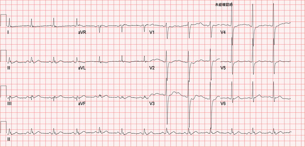
I found this case really interesting, and there's a lot to learn from it. So I pulled it ahead of the many other cases I want to write about.
A 73-year-old man came in in the middle of the night. Because the chest pain was severe, the ED doctor on shift also did a Chest CTA to rule out aortic dissection.
When I took over in the morning, my own habit is to take another look at the ECGs the previous shift did on the handed-off patients whose chief complaint was chest pain. The patient's initial TnI was 0.26 ng/mL, already >99th percentile of URL. A second set of cardiac enzymes was planned for later.
The initial ECG is shown above. After looking at it, I went straight to see the patient, to check how his chest pain was doing now.
Do you all see anything?
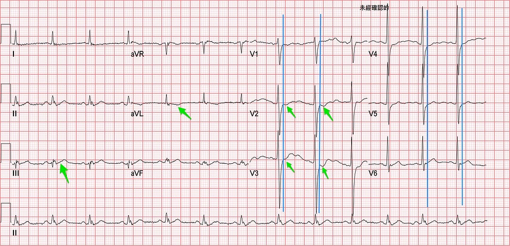
The vertical blue line is drawn at the J point. The J point is where you assess for STE or STD. The J point is the end of the QRS and the start of the ST segment. Above the baseline is STE; below the baseline is STD.
Using STEMI criteria means judging whether there's STE based on where the J point is, and only then can you diagnose STEMI. The most recent 2018 4th Universal Definition of Myocardial Infarction lays out clearly what an AMI is, and what can be called a STEMI.
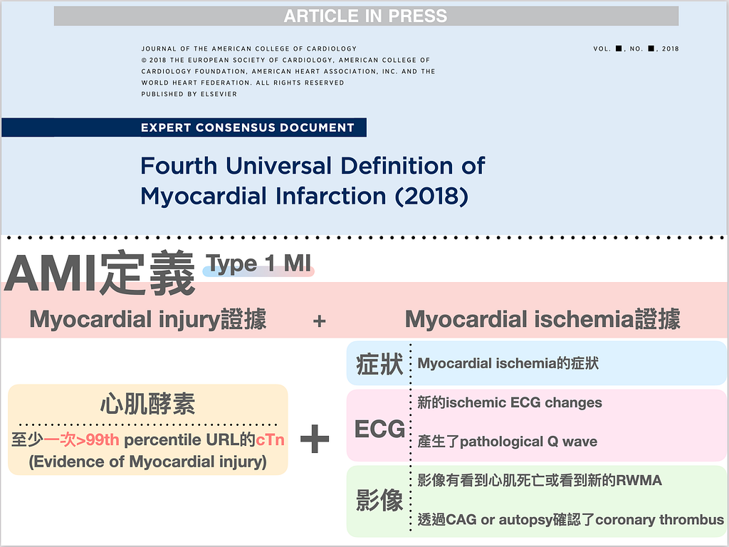
An AMI is evidence of myocardial injury (rising biomarker) + evidence of myocardial ischemia (symptoms or ECG or imaging abnormality). (Fig.2)
And to diagnose STEMI, you have to meet the STEMI criteria. (Fig.3)
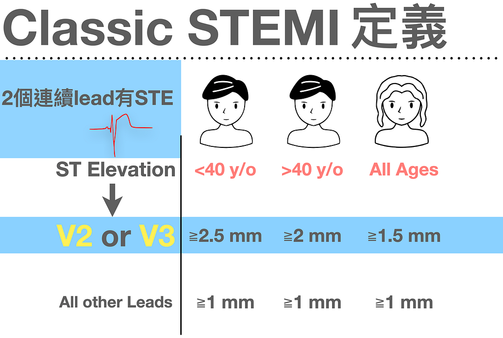
I have to stress this again and again: STE in V2/V3 is defined differently from the other leads. For V2/V3 you have to factor in sex/age together.
For example, in men <40 y/o, it has to be ≧2.5 mm in V2/V3 to count as meaningful elevation. And so on for the others.
This leads to a few interesting situations:
- It also means that if you grab any random person and do a 12 lead ECG, you're quite likely to see STE in V2/V3; whether it's meaningful elevation depends on the criteria above.
- If you don't see STE in V2/V3 (e.g., isoelectric baseline or minimal STD), you need to consider whether some force is pulling it down. Yep~~~ Post.OMI
- Following from that, how much minimal STD should make you consider Post.OMI?
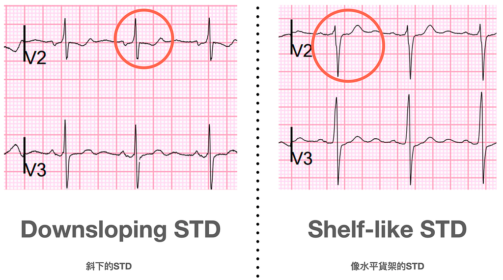
This post on Smith's ECG Blog says1 that any degree of STD in V2 or V3 must be considered posterior OMI until proven otherwise. Especially downsloping STD (per the above, even STD <0.5~1 mm deserves special caution)
The masters Ken Grauer and Smith have stressed the ECG morphology of V2 or V3 many times. Besides any degree of STD as above, if you see downsloping STD or shelf-like STD, watch out for Post.OMI.
In a post I wrote before this one, I went into more detail on how to diagnose Post.OMI2. In short: STDmaxV1–4(+) → points straight to a Post. wall MI until ruled out (in plain words: if there's STD in the precordial leads and the maximal STD is in one of V1-V4, consider a posterior MI).
Back to Case
In Fig.1, what I saw at the time was what looked like downsloping STD in V2/V3. The baseline wasn't very stable, but looking at V2 alone, all three beats seemed to show it.
Also, aVL had TWI and the ST segments in III/aVF were straight. Amal Mattu, when teaching, often calls this ST segment morphology the Checkmark sign (a tick mark). Normal STE is usually concave (upwardly concave); when the following appear, you have to consider an ischemic pattern of STE morphology. (Fig.5-A)
Also, the T waves in Lead II/III, relative to the QRS in front of them, count as HATW.

After that handoff, having seen this ECG, I thought all these leads showed ischemic ECG morphology, so I strongly suspected the patient had an OMI. But because the ECG didn't meet any STEMI criteria, it's very easy for this to get missed.
At the bedside, the patient said he still had chest pain. So I immediately got another ECG (Fig.6)
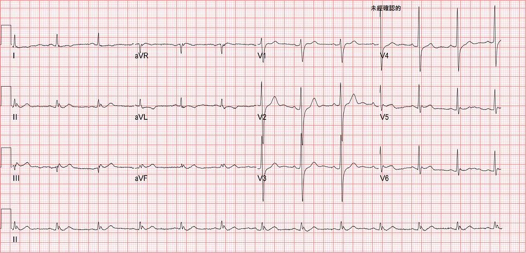
Fig.6 is the patient's second ECG, about three hours after arrival. The downsloping STD pattern in V2 or V3 was no longer there. But in Lead III/aVF the ST segments were still straight and the T waves were still HATW. The T wave in aVL was TWI. This ECG still didn't meet STEMI criteria, but it was absolutely an OMI.
To get more evidence of myocardial ischemia, I pulled out the Lumify again.
The PSAX papillary muscle view shows the lateral wall and inferior wall both moving poorly (RWMA+). (Fig.7)

This echo finding matched the ischemic territory on the ECG.
The patient had also had a Chest CTA on the previous shift to rule out aortic dissection.
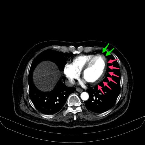
In Fig.8 you can see on the Chest CTA that the lateral wall of the LV, toward the more lateral part, has hypoperfusion (this is the myocardial perfusion defect we'll talk about below) (the darker red area). This also matched the ECG and echo findings.
Here we need to raise a question: could this hypoperfusion seen on non-ECG-gated contrast-enhanced CT be something else? Can it be trusted?
If you're interested, take a look at this paper3. Here's my summary of a few of its points:
- Cardiac CT (coronary CTA) can help us assess coronary artery stenosis, and can also assess myocardial perfusion defect (MPD) (i.e., an LV hypodense lesion like in Fig.8). But coronary CTA needs ECG gating, and also needs a high-end multislice MDCT.
- However, quite a few recent studies describe (see Fig.9) that ACS can be diagnosed on routine non-ECG-gated contrast-enhanced CT by detecting a myocardial perfusion defect.
- Although ECG-gated MDCT images all kinds of cardiac disease better, non-ECG-gated contrast-enhanced CT usually also provides enough information to identify some incidental cardiac abnormalities, such as a myocardial perfusion defect (MPD) that may be related to the patient's presenting symptoms.
- Cardiac motion artifacts are still the biggest factor affecting the diagnosis of ACS on non-ECG-gated contrast-enhanced CT
- A myocardial perfusion defect without myocardial ischemia is mainly due to cardiac motion artifacts (false positive), and the frequency of this is only 15–20%, which isn't high.
- Also, in ACS cases, because the AMI reduces regional myocardial wall motion, it also reduces cardiac motion artifacts and makes the myocardial border clearer (that's a bad thing and a good thing XD)
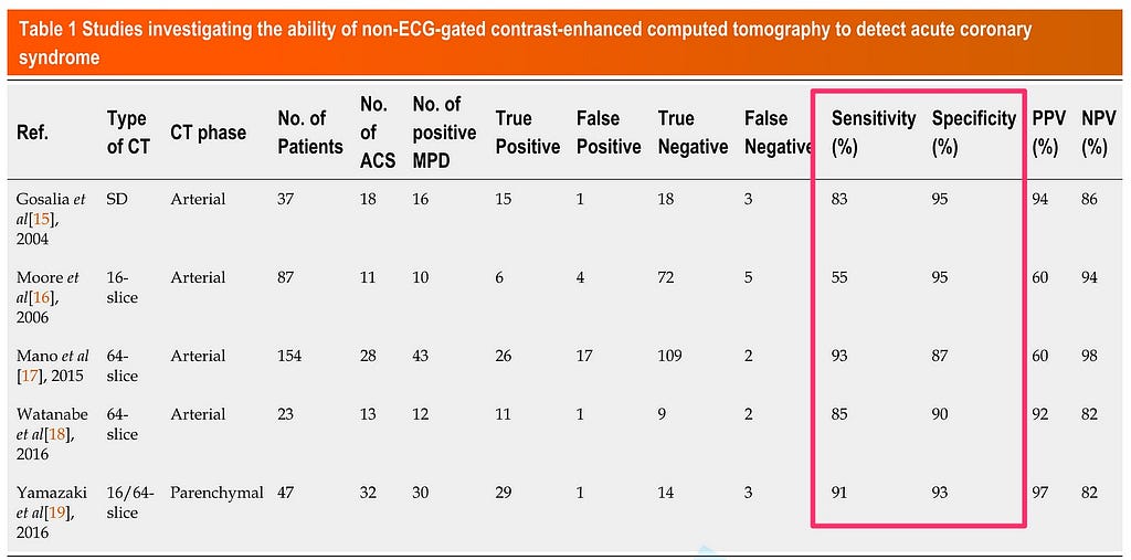
According to Fig.9, a myocardial perfusion defect (MPD) seen on non-gated-ECG contrast-enhanced CT has decent sensitivity and specificity for diagnosing ACS.
In short, this paper shows that to avoid missing treatment of a life-threatening condition like ACS, clinicians should be alert to the fact that even on routine non-ECG-gated contrast-enhanced CT, a myocardial perfusion defect is common when ACS is present.
Back to Case
At that point the evidence that the patient had an OMI kept piling up:
- ECG with HATW and straight ST segments in the inf. leads (Checkmark sign+), and downsloping STD in V2/V3 on the initial ECG
- Echo showing RWMA in the inf./lateral wall
- Myocardial perfusion defect on non-ECG-gated contrast enhanced CT
And all three fit a specific coronary artery territory.
On top of that, the patient still had ongoing chest pain after medical therapy. According to the ESC 2020 NSTEMI guideline, that makes him a patient with a very high risk feature4.
If we couldn't recognize that this patient had an OMI, then under the current STEMI/NSTEMI paradigm he should have been classified as NSTEMI (TnI⬆︎ plus evidence of myocardial ischemia). As I just said, refractory ischemic chest pain counts as a very high risk feature, so this patient should be scheduled for PCI within 2 hours to see whether the vessel is occluded (see Fig.10).
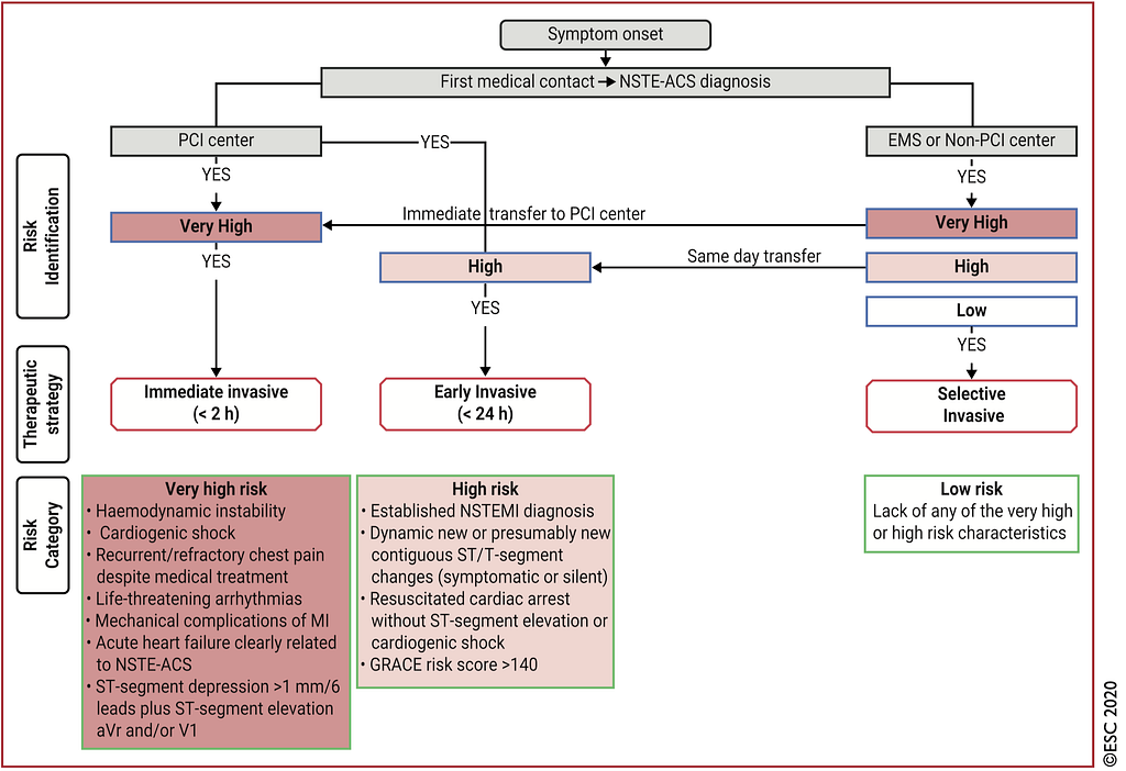
Fig.10 is probably the figure that shows up most often on my ECG blog XD
Note that the very high risk features in Fig.10 say almost nothing about how the ECG has to change before you do emergent PCI (the only exception mentioned is aVR STE + STD in ≧ 6 leads). In other words, if the patient has recurrent chest pain that doesn't respond to treatment, you should call the CV man to evaluate for cath, rather than doing ECG after ECG to see whether it turns into STE.
Of course, if serial ECGs show STE, great, because the CV man will definitely buy it and go in.
But…. it's exactly this but…..
Last month Smith's ECG Blog had a post5 comparing the STEMI/NSTEMI paradigm with the OMI/NOMI paradigm, and one of its points was about serial ECGs.
Let me list the key points: (no, ChatGPT didn't write this)
- In the STEMI/NSTEMI paradigm, when a patient has ischemic symptoms and the ECG doesn't meet STEMI criteria, serial F/U ECGs are done ➡ but STEMI criteria aren't very sensitive for ACO (meaning that when a patient's artery is occluded, it doesn't often show up as STE; more often it shows up as a non-STE ECG pattern)
- STEMI(-)/OMI(+) patients may get many ECGs that repeatedly show the diagnosis of OMI (just like my case) but repeatedly fail to reach STEMI criteria ➡ usually these serial F/U ECGs don't evolve to meet STEMI criteria, and they only change after the best window for intervention has passed (damn!!! so sneaky XD)
- Serial ECGs looking for STEMI criteria may keep missing STEMI(-)/OMI(+), leading to delayed PCI
So serial F/U ECGs that try to catch the appearance of STE may delay PCI for this patient, because STEMI(-)/OMI(+) often may not evolve into STEMI(+)/OMI(+).
So as long as it's NSTEMI + a very high risk feature → please call the CV man
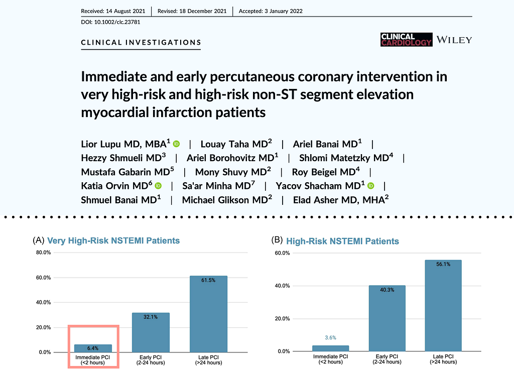
Although we know that the proportion of NSTEMI patients with a very high risk feature who actually get taken for PCI within <2 hours is low. It's like this all over the world (Fig.11).
But don't get discouraged; there will always be times you manage to help the patient
Back to this case~~
So I called the CV man myself and told them my concerns. The CV man agreed with me and was willing to take the patient to cath right away.
I typed the last words into the electronic chart, and right after that the patient went up for cath.
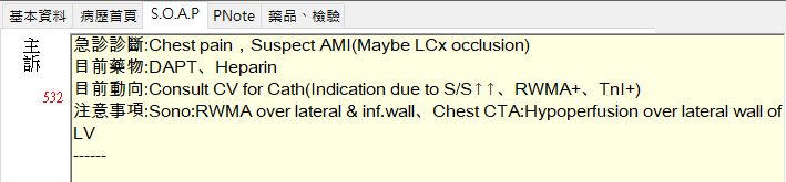
CAG report: LCx total occlusion➡CAD with TVD s/p CAG, with LAD 50–60% occlusion, RCA: CTO, LCX total occlusion, DES*1 over LCX
Key points:
- How to diagnose classic STEMI (V2/V3 defined differently)
- Definition of AMI (Fig.2)
- Is minimal STD or an isoelectric baseline in V2/V3 meaningful?
- Ischemic-pattern STD (downsloping/shelf-like STD)
- Can a myocardial perfusion defect (MPD) seen on non-ECG-gated contrast-enhanced CT be trusted?
- STEMI(-)/OMI(+) usually doesn't evolve into STEMI(+)/OMI(+) that meets STEMI criteria
- Know which situations count as very high risk features in NSTEMI
- If you're worried the patient has an OMI but there's no ECG meeting STEMI criteria, look for more evidence of OMI (serial F/U ECG, TnI, echo). Call the CV man yourself and tell them what you think; it may help the patient get PCI sooner and lose fewer heart cells.
Additional references: 1 2 3 4 5
Dr. Smith’s ECG Blog: Any ST depression in V2 and V3 is posterior OMI until proven otherwise, especially if downsloping — link ↩︎ ↩︎
[This is a STEMI, do you believe it? A case from my night shift yesterday: a 50-year-old man who used to catch glass eels.] (in Chinese) | by Tsao Jian-hsiung | Medium — link ↩︎ ↩︎
Yoshihara, S. (2022). Acute coronary syndrome on non-electrocardiogram-gated contrast-enhanced computed tomography. World Journal of Radiology, 14(2), 30–46. https://doi.org/10.4329/wjr.v14.i2.30 ↩︎ ↩︎
Collet, J.-P., Thiele, H., Barbato, E., Barthélémy, O., Bauersachs, J., Bhatt, D. L., Dendale, P., Dorobantu, M., Edvardsen, T., Folliguet, T., Gale, C. P., Gilard, M., Jobs, A., Jüni, P., Lambrinou, E., Lewis, B. S., Mehilli, J., Meliga, E., Merkely, B., … ESC Scientific Document Group. (2021). 2020 ESC Guidelines for the management of acute coronary syndromes in patients presenting without persistent ST-segment elevation. European Heart Journal, 42(14), 1289–1367. https://doi.org/10.1093/eurheartj/ehaa575 ↩︎ ↩︎
Dr. Smith’s ECG Blog: 50 year old with acute chest pain, with ‘normal’ ECG and falling troponin — link ↩︎ ↩︎



Thoughts on this case? Leave a comment, a like, or a reaction below.
comments powered by Disqus