Translated from the original Traditional Chinese post. Read the Chinese original →
A case of out-of-hospital cardiac arrest — what was the cause?
76-year-old woman.
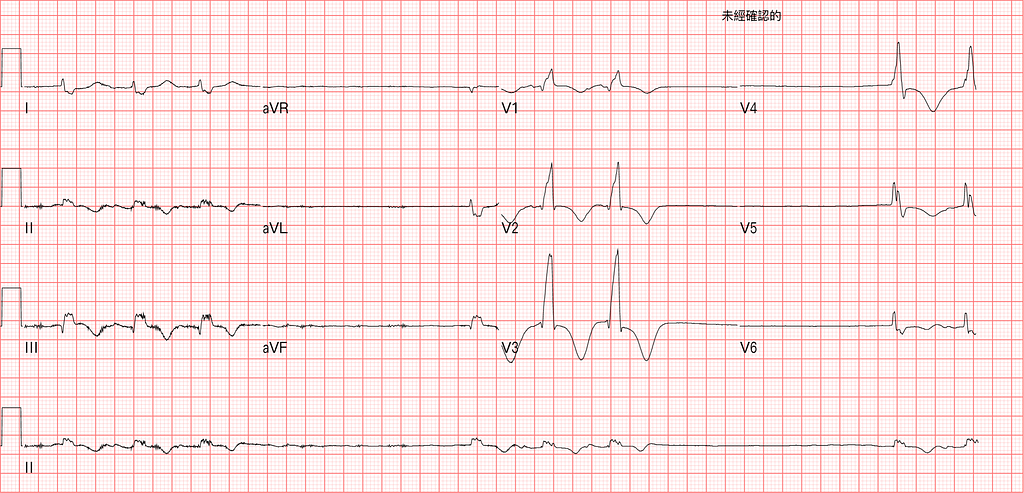
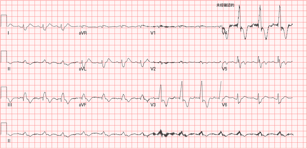
Arrived at an outside hospital in out-of-hospital cardiac arrest; after resuscitation, she got ROSC. She was transferred to our hospital, and the first ECG on arrival in our ED is shown above (at this point she was about to crash). Her heart stopped very soon after. After roughly another 10 minutes of CPR, she got ROSC again. We did another 12-lead ECG (Fig 2).
What do you think caused this?
First look at the ECG on arrival, right before she arrested. Long pause, RAD, RBBB, TWI over V1~V6, II, III.
Then look at Fig 2 → Rate: 90 bpm, SR, RAD, RBBB, TWI over V2~V4, II, III, aVF.
Here we need to think through and clarify a few questions!
- When there's TWI in V1~V4, what do you need to consider?
- Does the Precordial lead TWI here need to make us consider Wellens' syndrome?
- When the diagnosis comes down to Wellens' syndrome vs. PE, how do you make the differential diagnosis? What V2~V3 morphology leans toward RV strain?
When there's TWI in V1~V4, consider the following:
- Post.MI → when the maximal ST depression (possibly with TWI) is in V1~V4, think Post.MI first (reflexively check the Inf. wall for STEMI).
- RBBB normally has STD and TWI in V1~V4, which represents appropriate discordance → what does that mean? It means the ST segment and T wave point in the opposite direction from the QRS of abnormal conduction (abnormal conduction caused by the BBB)
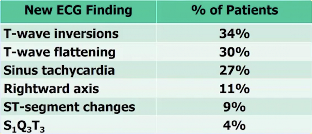
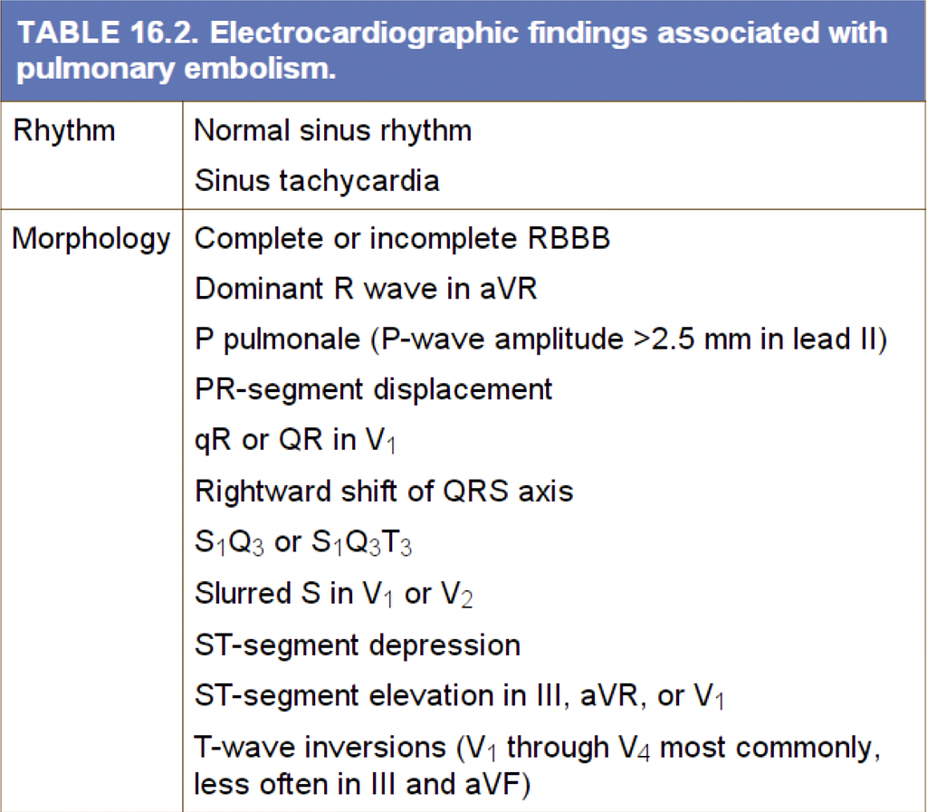
- TWI in the R’t precordial leads means you need to put PE on the differential. The most common ECG finding in PE is T wave abnormality, including flattening and TWI (about 60% in total). Also, New onset RBBB/ICRBBB should make you watch out for PE.
RBBB/ICRBBB appears → because the RBB runs along the septal wall next to the RV, and the RV wall isn't thick, RV strain causes RV wall ischemia, which in turn produces RBBB/ICRBBB
- Wellens' syndrome also belongs on the differential. It's defined as Biphasic T waves or Deeply TWI in V2/V3 (sometimes it can extend to V1~V6). With Wellens, you need to pay attention to its definition — pay attention to what? I'll cover that next time when I have time~
Does the Precordial lead TWI here need to make us consider Wellens' syndrome?
Yes, Wellens' syndrome does need to be considered
When the diagnosis comes down to Wellens' syndrome vs. PE, how do you make the differential diagnosis?
First, when the diagnosis comes down to Wellens vs. PE, it means we think this patient may have an AMI or a PE. So why do we think it's AMI (Wellens) or PE? Because we saw TWI in the R’t precordial leads, and that's what put the idea in our heads.
Here are some differentiating tips that can make our DDx more precise
First, we need to know that PE causes Pul.HTN, which in turn causes RV strain. So which ECG findings suggest there may be RV strain? (Four ECG signs of RV strain → note that PE, COPD, Acute Pul.HTN, and hypoxia from Pneumonia can all produce these changes: the ECG can't diagnose the underlying cause)
- Tall R wave in V1(R/S >1) → only present as a normal variant in 1%, so it's not common in the ED — memorize the table below!
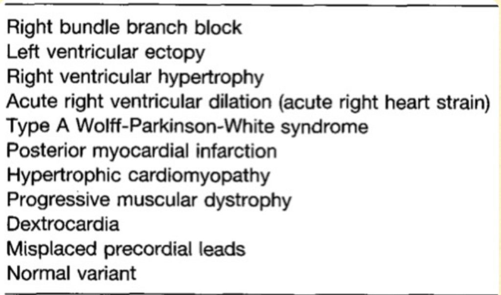
- RAD
- ST changes (especially STE in the Rightward leads: V1, V2, aVR, III)
- TWI over ant.septal ± Inf.lead
Here's a very important Paper conclusion: if the diagnosis is down to ACS vs. PE, when TWI is in the precordial leads and also in Lead III → Favor PE, not ACS (TWI in III and V1 is present in 1% of ACS and 88% of PE, so 99% specificity)²
After all that? How do you actually tell the TWI of PE from Wellens' T waves?
- Wellens' T waves appear in the pain-free period
- ACS, or especially reperfusion (Wellens), rarely has tachycardia
- Wellens' T waves and PE's T waves look different (sounds kind of mystical LOL)

Look closely: the top row is all different PE cases, and the bottom row is all different Wellens cases. Notice anything different?
What V2~V3 ECG morphology leans toward RV strain? (If RBBB is present, the criteria below don't apply)¹
- Usually a bigger S wave than R wave
- Usually an isoelectric J point or elevated J point, followed by upwardly convex STE and then TWI (meaning PE's TWI looks different from the TWI of Wellens' waves)
PE's TWI is convex STE that then bends downward like a roller coaster; ACS-Wellens TWI drops steeply and is sharper
- Usually some STD in V4~6, and the inf. leads will also have TWI
Back to the original Case~~~
This patient's ECG showed RAD, RBBB, TWI over V2~V4
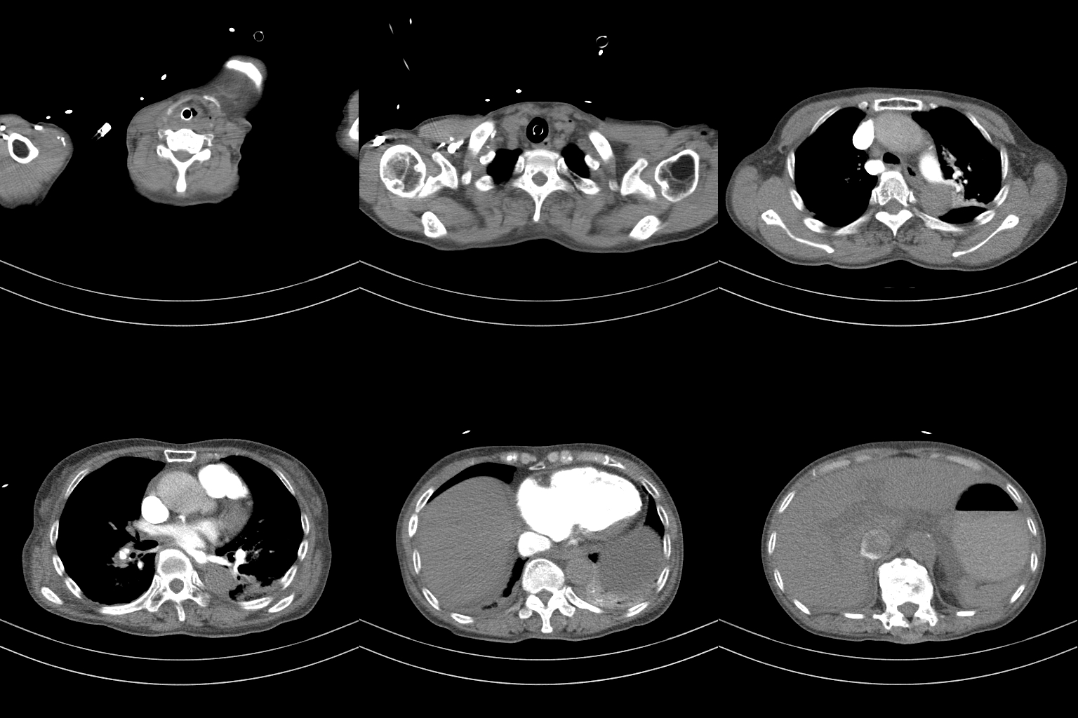
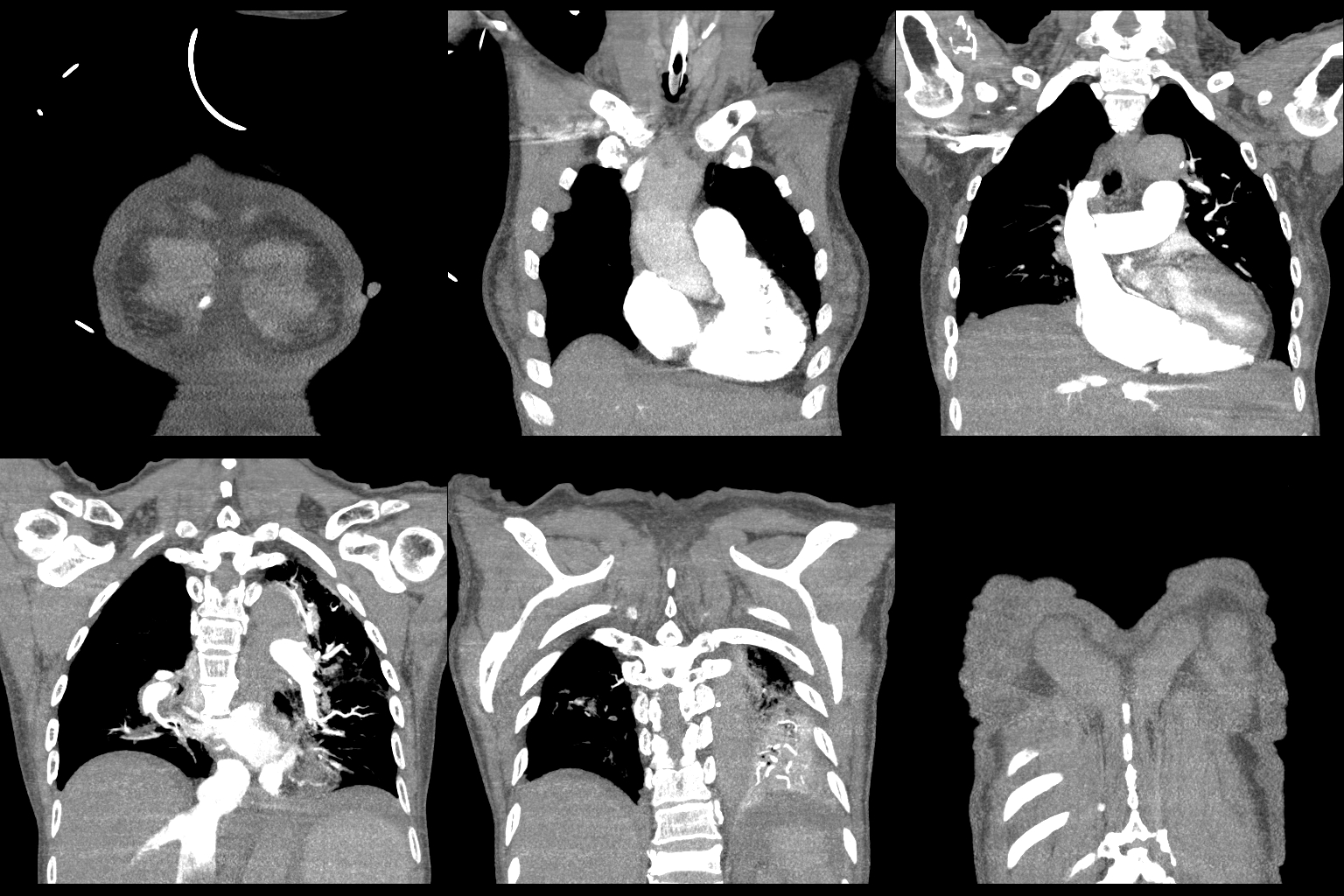
We then did a Chest CTA, which showed the right upper pulmonary vessels completely occluded.
The patient went home to heaven that afternoon………
- Dr. Smith’s ECG Blog: A crashing patient with an abnormal ECG that you must recognize — link
- Electrocardiographic differentiation between acute pulmonary embolism and acute coronary syndromes on the basis of negative T waves



Thoughts on this case? Leave a comment, a like, or a reaction below.
comments powered by Disqus